The process of preparing the mother's body for birth resolution occurs all 9 months of gestation. For an easier and more comfortable appearance of the baby, the woman's pelvic bones also expand slightly. This process is natural for everyone. However, after giving birth, a woman realizes that with her usual weight, her hips have become wider. How does the recovery of the pelvis after childbirth occur, and does it occur at all?
The female body is a well-established system for bearing a child. Throughout pregnancy, a woman undergoes multiple changes, both external and hormonal. However, the most visible and tangible physiological changes await mom on last dates, immediately a month before the appearance of the baby.
Changes occur at the hormonal level. If earlier the body spends all its forces on the development of the fetus, now reserve forces are spent on preparing all internal organs to childbirth. The bones of a woman do not remain unchanged. The body pays a special role to softening the pelvic bones.
During prenatal natural preparation, the pelvic bones (partly under the influence of hormones) become more elastic and flexible. This is necessary so that the bones of the baby's skull pass more smoothly, and the woman's birth canal is able to release the baby into the world.
Naturally, it should be assumed that the reverse "transformation" of the pelvic bones cannot occur in a few weeks, and therefore the young mother should be patient. Many young mothers worry about the fact that their sizes will never be the same, but such fears are only fears.
How long does it take to restore the pelvic bones after childbirth?
Pregnancy and childbirth, for sure, for every woman is the most crucial period in life. During this time, the girl's body learns to be a mother, to work not only "for itself", but also for the benefit of another body - a baby. The mother's body courageously endures all the trials, pains and changes that are necessary for just one single day - childbirth.
Expansion of the pelvic bones is almost always accompanied by aching and dull pains in the lumbar region, pelvis, thighs, and womb. However, the actual expansion of the bone in this place cannot exceed 2.5 cm. Despite such small numbers, the woman’s pelvis and its width, the difference between “before” childbirth and “after”, feels immediately after the birth of the child.
It takes at least 3 months to fully restore the previous size. As many reviews of the mothers themselves say, the pelvis completely converges to its previous size no earlier than in the second, third month after the birth of the baby. At the same time, pain, discomfort or aching manifestations in the pelvic region are not observed.
See also recovery. female body after childbirth in our article
Pregnancy is a special condition of a woman, which is accompanied by changes in the hormonal background. All changes are aimed at adapting the body to childbearing and childbirth. However, due to some features of the woman's body or due to the fact that the concentration of hormones increases several times, various painful sensations may appear.
These are pelvic and lumbar pain that occurs in 25-50% of pregnant women and women in the postpartum period. Most often they are affected in the last months of gestation. This is due to the fact that the fetus becomes quite heavy, and the concentration of certain hormones in the blood is at a maximum.

An important point is that the concentration of some hormones increases, while others decrease during different periods of gestation. In this regard, the severity of their action on the fetus and the body of a pregnant woman changes.
Let's see why the pelvic bones, lower back and sacrum hurt during pregnancy and after childbirth? Is this pain normal after childbirth?
Physiological action
It is believed that the hormone relaxin weakens the ligaments and helps soften the cartilage of the pubic and iliosacral joints. Its concentration increases at the end of the third trimester, thereby preparing the birth canal. Under the influence of relaxin, the cartilaginous disc of the pubic articulation, interosseous, dorsal, ventral sacroiliac ligaments of a similar articulation fall. During pregnancy and in the first days after childbirth, the pelvic bones hurt most intensely, it may be difficult for a woman to lie on her side and back. The pain syndrome is localized in the region of the sacrum, lower back, and hip joint. After childbirth, the condition returns to normal within a few weeks, the pain subsides.

excessive action
With an increased concentration of the hormone relaxin and its metabolites in the blood or with high sensitivity to it, it can cause excessive relaxation of the pelvic ligaments and cartilage. With pathological relaxation of the cartilage of the pubic joint, the pelvic bones diverge and symphysitis occurs, and with the defeat of the sacroiliac - sacroileopathy.
These diseases are accompanied by pain in the bones of the pelvis, sacrum, coccyx and the area of the hip joint. Pain of moderate or moderate intensity, increases with palpation in the pubic area and when getting out of bed. Patients complain that the pelvis and its bones hurt when going to the toilet. After rest, pain usually disappears and worsens with any physical activity.
Injuries during childbirth
Increased activity of relaxin, large fetus, complications of labor activity can lead to rupture of the pubic symphysis or damage to the coccyx. With a rupture of the pubic joint, the pelvic bones diverge in this area up to 5-7 cm and immediately after childbirth cause pain of medium or high intensity. The pain is exacerbated by any movement, and in bed after childbirth, the puerperal is in a forced position - the “frog position”.
With an injury to the coccyx, the puerperal will feel pain only when she gets on her feet or sits for a very long time, during the act of defecation. The pain is intense, pulling in nature, aggravated by getting up from a sitting position, bending forward, tension of the muscles of the pelvic floor. The injury may be accompanied by a curvature of the posture and spine - antalgic posture.
- objective data.
In addition to collecting complaints of pain in the relevant area, studying the anamnesis, the doctor conducts an examination and palpation to find out the distance between the pelvic bones, to assess the function of the joints. Also, with a rupture of the pubic joint or symphysitis, the patient will not be able to raise her legs in an unbent position upwards while lying on a hard couch. There may be difficulty in moving up the stairs, a change in gait, which are diagnostic criteria for making this diagnosis.
- Radiography.
The main research method, which is a litmus test in the diagnosis of postpartum injuries and lesions of the ligamentous apparatus of the pelvis, remains radiography. It is thanks to her that it is possible to make such diagnoses as "symphysitis", "rupture of the joints", "fracture and dislocation of the coccyx", "sacroileitis".

According to x-ray data, 3 stages, or degrees of severity, of symphysitis are distinguished:
- Stage I - the distance between the pelvic bones is from 0.5 cm to 1 cm;
- Stage II - from 1 cm to 1.9 cm;
- Stage III - more than 2 cm.
With an increase in the distance of more than 2-3 cm, it is worth thinking about breaking the pubic joint.

Treatment of diseases associated with damage to the ligamentous apparatus of the pelvis after childbirth is aimed at creating favorable conditions for healing and strengthening the surrounding ligaments. For this purpose, postpartum women are prescribed orthopedic pillows in the form of the letter C, seat cushions in the form of a ring (donut), massage, water aerobics and swimming.
A good assistant in the treatment is a bandage, which provides a quick and effective strengthening of the ligaments, restoration of the function of the musculoskeletal system, with its simultaneous unloading. Wear a bandage throughout the disease. There are also special corsets that ensure the immobility of the coccyx and contribute to its healing. They also prescribe medications that are aimed at reducing inflammation, relieving pain - paracetamol, ibuprofen, B vitamins.

Timely access to a doctor with complaints, a detailed history and characteristics of pain (where and how it hurts) allows you to early stages diseases to carry out as much as possible effective treatment, is a protection against the development of complications.
Sources:
- Obstetrics / V.I. Duda - Minsk - 2013 - 576 pages.
- Obstetrics and gynecology T.1 / V.M. Zaporazhan - 2005 - 472 pages
- Obstetrics. National allowance / E.K. Ailamazyan, V.I. Kulakova, V.E. Radzinsky, G.M. Savelyeva - 2009 - 1200 pages.
After the birth of a baby, women are often accompanied by a number of problems and troubles. Many diagnoses can be made to a girl who has experienced similar changes at the physiological and psychological levels. Symphysitis is a divergence of the pelvic bones after childbirth. The concept includes damage and changes in the pubic joint. This phenomenon occurs after changes in the girl's body.
Expansion of tissues is formed in several steps: the pelvic bones loosen, soften, swell, stretch, expand, diverge. After a break occurs and begins inflammatory process, which is called symphysis.
The hips can become wider after and already during the bearing of the child. This problem appears due to changes occurring in the body of a woman in order to facilitate the process of delivery. This is a natural reaction of the body when the hips become wider after that.
The body releases a substance called relaxin. Its function is to relax, increase the mobility of the joints, the distance between the femoral joints. An increase in pubic articulation is 5-6 mm wide. Over time, everything takes its usual position: the cartilage becomes denser, the ligaments become more elastic, and the joint space acquires its former width.
Symptoms of the disease
The pelvis became wider after due to the weakening of the pubic bone. Signs of symphysiopathy can appear even before the birth of a child. In the case when a girl does not receive enough trace elements and minerals. This leads to calcium starvation, which provokes the occurrence of layering of the nails, frequent convulsions, tooth decay, and fatigue. The disease is especially characterized by frequent pain, redness and swelling. Movement may be disturbed and signs of fever may appear.
During the divergence of the pelvis, wandering pains often appear. Such symptoms can be mistakenly confused with the diagnosis of osteochondrosis and sciatica.
If the connections diverge during pregnancy, there is a threat of interruption.
When do tissues begin to diverge during pregnancy? Unpleasant sensations appear during the 2nd-3rd trimester: the pain reminds of itself when walking or standing for a long time. Such conditions are encountered by specialists very often. At what time the pains begin to disturb, it is impossible to give a definite answer. The disease can be latent.
In the second half of the period of bearing a child, the abdominal muscles are tense to hold back the pubic joints. natural way. After that, the pelvic bones do not immediately converge and the abdominal press is initially quite flabby.
Symphysiopathy is a fairly strong relaxation of the pubic joint. It is one of the manifestations of toxicosis characteristic of this period, with damage to the system of tissues and joints. The diagnosis becomes chronic. Symphysiopathy does not apply to physiological processes when the bones temporarily parted during childbirth.

Degrees and causes of symphysis
There are several degrees of expansion of the pelvic bones after childbirth:
- The first degree is from 5 to 9 mm.
- The second degree is from 10 to 20 mm.
- The third degree is more than 20 mm.
The occurrence of symptoms of the disease indicates a violation of the metabolism of calcium and phosphorus in the body and a deficiency of vitamin D group (a considerable amount is found in butter, sea fish, egg yolks, cheese, caviar, mushrooms and liver). As a result, thyroid diseases are formed, intestinal function is disrupted, there is a risk of diabetes and kidney failure.
Ailments of this nature make themselves felt with poor nutrition, devoid of nutritional value and essential substances, or with severe toxicosis, when all the benefits of food do not have time to come in sufficient measure.
Diagnosis
When the pelvic bones begin to diverge during pregnancy, the sensations are rather unpleasant. Now everything is wider than usual, and this causes severe symptoms. The malaise is aggravated during attempts to walk, move, or change position. In order to alleviate discomfort, the girl intuitively assumes the “frog” position: lying on her back, she bends her knees and turns her hips.
A characteristic symptom of when the pelvis is too dispersed after childbirth will be a slow, pumping from side to side, gait. It can be observed even during the period of bearing a baby. The doctor can only clarify the diagnosis with a thorough examination of the damaged area. Palpation will help to determine the degree of soreness on both sides. The measure of deepening is determined by pressing with a fingertip.
Effective diagnostic methods:
- Ultrasound. The method is characterized by increased safety: it does not harm the mother and child. Provides complete and accurate study.
- X-ray. This method is able to detect only stretching, the presence of pathologies is not determined. X-rays harm the baby.
- Analyzes. The results will help to detect the degree of presence in the blood and urine of the concentration of potassium and magnesium.
These manipulations are able to predict the problem if there are no complaints and recognizable symptoms.

Treatment
With proper and timely treatment, surgery can often be avoided.
During a mild expansion, when the recovery of the pelvis after childbirth occurs quickly, doctors strongly recommend:
- limit any physical activity;
- wear a tight bandage;
- sleep on a special mattress;
- take fish oil capsules and preparations rich in calcium, magnesium;
- if necessary, take painkillers in capsules, suppositories, tablets and ointments.
If the situation is more serious:
- the main task is to achieve convergence of the pubic joints and keep them in the desired position;
- strict bed rest is required for a period of 2-6 weeks;
- bandaging and tight bandaging will help;
- physiotherapy and cold compresses are prescribed;
- additionally taken anti-inflammatory, antibacterial and anesthetic drugs;
- It is recommended to follow a special diet high in calcium.
Preventive measures
In order not to think about when the pelvic bones will begin to diverge during pregnancy and in order to avoid damage to the pubic joint in the future, it will be useful to adhere to certain rules.
- Follow a balanced diet rich in all minerals. Eat enough food that contains manganese, chlorine, calcium, magnesium, sodium and phosphorus. Sources: yogurts, sour cream, cheeses, cottage cheese, milk, eggs (especially yolk), rabbit meat, chicken, lean veal, fish and shrimp, caviar, legumes (peas, beans, chickpeas, corn), nuts (almonds and cashews) , greens and mushrooms.
- Ensure yourself sufficient activity: engage in light sports, attend specialized courses, walk a lot.
- visit fresh air.
- Take the right pills and supplements in addition to proper nutrition.
- Regularly visit a gynecologist and follow all the recommendations of doctors.

When everything is back to normal
Pain, discomfort, an increase in volume - regular complaints of women who are expecting a baby and worry that their hips have become wider in size. Stretching and softening of tissues is a natural process for easy crumbs to come out. This phenomenon is temporary. Of course, a couple of days will not be enough for everything to fall into place.
It is difficult to say how long after childbirth and how long the hip joints narrow back. In a significant part of cases, after the birth of the crumbs and the completion of the breastfeeding process, the hormonal background is completely restored.
After childbirth, the bones converge quickly with:
- active lifestyle;
- proper nutrition.
The resulting fat deposits will go away, the connections will take their usual position and the appearance of the girl will acquire its former contours. To speed up the process, some are willing to go under the surgeon's knife. This is an erroneous opinion, because the body still needs proper time to recover from the stress experienced. He himself will return to normal when necessary, and after that the body will no longer be wider.
Childbirth is a complex process involving many body systems. After the birth of a child, a woman in labor feels weakness, fatigue, and sometimes pain. So, in 50% of women, the pelvic bones hurt after childbirth, which gives them discomfort, prevents them from living fully and caring for a child.
Causes of pain
Causes discomfort several in the pelvic bones. They are associated with changes that allow the body to adapt to pregnancy and ensure the passage of the child through the birth canal.


Pulling or sharp pain in the pelvic area with lumbago is a reason to consult a doctor for an examination and treatment.
Possible diseases
Many pregnant women experience a disease such as symphysitis. The pubic bones are connected to each other by a fibrocartilaginous disc - the symphysis. Under the influence of hormones and fetal pressure, the symphysis is stretched by 5–6 mm or more. As this discrepancy increases and inflammation joins, symphysitis is diagnosed.
Symphysitis is characterized by pain when walking, changing body position, physical activity. The gait changes (becomes similar to a duck), there is discomfort during urination and defecation.

The disease is diagnosed by palpation. When pressing on the symphysis in the pubic region, acute pain occurs. Ultrasound is also used for diagnosis, which, however, allows small errors. The most accurate degree of symphysitis allows you to establish an X-ray examination. If the discrepancy is more than 1 cm, doctors may decide to refer to a caesarean section.
Rupture of the symphysis during childbirth is a rare phenomenon that requires surgical intervention and prolonged, for several months, bed rest.
Postpartum recovery includes a number of measures that allow you to connect the dispersed parts of the pubic joint and limit their movement:

- Bed rest and reduced physical activity. The first time after childbirth, women need to reduce the load, if possible, entrusting the care of the child to relatives.
- Bandage. Special bandages have been developed that wrap around the hips, limiting the mobility of the hip joints and pubic bones.
- Visiting an osteopath As the condition improves, the doctor will prescribe a set of physical therapy exercises.
- Restoration of vitamin and mineral balance to strengthen bones and connective tissue.
- Anti-inflammatory therapy and physiotherapy in a day hospital.
The course of treatment is compiled individually after examination and consultation of the therapist, gynecologist, osteopath and surgeon. In some cases, an operation is prescribed with the introduction of steel supporting structures.
How to relieve pain
 Severe pain in the coccyx and pubic area is relieved by painkillers, conditionally allowed for breastfeeding: Ibuprofen, Paracetamol. With artificial feeding, you can take any effective painkillers to which the woman in labor has no individual contraindications: Pentalgin, Ketanov, No-shpa, etc.
It happens that the transferred stress and unstable hormonal background depress the central nervous system and change the perception of pain. In this case, it is recommended to take sedatives that are relatively safe for breastfeeding: Glycine, Valerian, Motherwort tablets.
Preventive measures

Prevention of symphysitis during pregnancy avoids postpartum complications.
- It is recommended to do special gymnastics, stretching and strengthening the muscles of the pelvic floor.
- With an uncomplicated pregnancy, you need to walk a lot, walk in the fresh air. Walking helps to strengthen the musculoskeletal system, and ultra-violet rays increase the production of vitamin D, necessary for the absorption of calcium.
- It is necessary to take a vitamin-mineral complex, which includes calcium and phosphorus, which help strengthen bones and connective tissue.
- Proper nutrition, combined with moderate physical activity, helps maintain a normal balance of nutrients, ensuring good health and functioning of the body systems that will be involved in childbirth.
Many pregnant women and women who have given birth have pain in the pelvic bones. This is due to the changed hormonal background, increased load and is a variant of the norm. Attention to yourself, observation of sensations, regular examinations and following the doctor's recommendations will help to avoid injuries and quickly recover after the birth of a child.
The main functions of the pelvic bones include:
- protective - prevention of possible injuries to some vital organs localized in the area under consideration;
- the pelvic bones are involved in the process of blood formation, this is facilitated by the presence of red bone marrow in them;
- motor - a healthy pelvis becomes the key to harmonious, natural movement of the body;
- the bones of the pelvis hold the column of the spine and are responsible for a straight, correct posture.
Causes of pelvic tilt
One of the most common diseases of the pelvis is the displacement of its bones. The pelvic tilt develops under the influence of factors:
- Sedentary lifestyle (lack of exercise)- Frequent sitting in a sitting position leads to the weakening of some muscles and contributes to the constant tension of others. This phenomenon does not pass without a trace, being the cause of an oblique pelvis.
- Lifting heavy objects- weightlifting without proper instruction is not recommended. Incorrect physical exercises (jerks, lifting and carrying dumbbells with one hand, etc.) lead to the development of the pathology in question.
- Various injuries of the pelvic bones- Injuries usually appear as a result of blows, falls, bruises. Of particular danger are open fractures of bones, rupture of the pelvic ring.
- Pregnancy status- the structure of the female pelvis is ideal for childbirth, but during gestation, the bones of this area often shift.
- Pelvic muscle injury- damaged muscles become less elastic, are constantly in a tense state. The direction of displacement depends on which muscle is damaged. The defeat of the lumbar muscles leads to the displacement of the pelvis forward, and the injury of the quadriceps muscle leads to a modification (flexion) of the hips.
- Herniated disc- the development of pathology in this case is due to chronic muscle spasm.
- leg length difference- when one limb in a person from birth or due to an illness is shorter than the other, then the pelvis twists or shifts from right to left, rarely from back to front.
- congenital or acquired scoliosis.
- Transferred surgery in the region of the pelvic or iliac bones.
Symptoms of the disease
Some symptoms of the disease can be recognized on early stages development. It is necessary to take a closer look at the features of the course of the disease in a timely manner and seek the help of a specialist for further treatment:
- Severe pain that manifests itself during walking, running, leg movements.
- Unsteady gait, unreasonable frequent falls, loss of balance.
- There is pain in the back, lower back, neck, shoulders. The pain gradually radiates to the legs.
- Unpleasant sensations in the genital area, groin.
- Dysfunction of the genital organs (in men, against this background, there is a sharp decrease in libido).
- Pain in the buttocks, thighs.
- One limb becomes gradually longer than the other.
- Bladder dysfunction.
- Diseases of the gastrointestinal tract (intestinal disorders).
- Pain in the knees, ankles.
- Movement becomes stiff.
- One shoulder will be higher than the other (the person unconsciously raises it).
If several signs of the disease are found, it is necessary to visit a doctor and think about the rational treatment of a distorted pelvis.
Diagnostics
You can determine the disease yourself, but to complete the clinical picture, you need to visit an orthopedist-traumatologist. The doctor begins the examination of the patient after a detailed anamnestic analysis. It is important to identify the root cause that provoked the development of pathology, and then proceed to treatment. Having completed the collection of anamnesis, the specialist will conduct a visual examination of the patient, probe the damaged, disturbing areas.
Delivery of an accurate diagnosis is impossible without an instrumental medical examination. The most effective methods of such diagnostics include:
- X-ray examination of the spine and pelvic bones of the patient.
- Magnetic resonance imaging, which will reveal possible curvature, damage in the spine or pelvic bones.
- Tomography performed using computer technology.
Treatment of pelvic tilt
For effective treatment of displaced pelvic bones, it is necessary to identify the root cause of the disease. The fight against the disease is to eliminate the factors that contribute to its development. Drug therapy in the treatment of pathology is based on taking the following medications:
- Anti-inflammatory drugs (non-steroidal).
- If pain is often disturbing, then it is advisable to use various analgesics.
- It is recommended to apply healing ointments to the damaged area of the body.
If symptoms of a twisted pelvis are detected, treatment for this disease should begin as early as possible. The neglected form of pathology requires surgical intervention, therefore it is better to treat the disease in the early stages. In Moscow and other large cities of Russia there are specialized clinics where effective complex treatment is carried out with an oblique pelvis.
There are other methods of treating pelvic tilt, which should be consulted after a medical consultation:
- Therapeutic exercise, performing various gymnastic exercises.
- Manual therapy, massage of the damaged area of the body.
- Physiotherapy procedures.
Distortion of the pelvis, the treatment of which is effective only in the first stages of development, is not always subject to complete healing. But an integrated approach to treatment will give the expected positive result.
Twisted pelvis in children
When the pelvis is tilted in a child, treatment should be urgent. Wellness procedures in this case require more time. Among the reasons for the displacement of the pelvic bones in babies, congenital pathology is distinguished. But most often the disease is acquired. Children spend a lot of time at their desks at school, and then at home in front of computer monitors and TVs. Low physical activity leads to the development of the disease in question.
For the treatment of children's oblique pelvis, it is important to normalize the deformed parts of the body.
It is worth strengthening the child's back muscles, leveling his posture with the help of a variety of physiotherapy procedures (mud therapy, hydrotherapy, massage treatment, exercise).
Rehabilitation exercises as a means of combating the disease
Treatment with physical culture is a fairly effective remedy against the developing skew of the pelvic bones. Most experts recommend the following exercises to patients:
- Lie on the floor, place your arms along the body, substitute a soft pillow under your feet. Relax all the muscles and start doing rolls on the stomach to the right and left. It is not recommended to tear off the hips from the floor; for greater efficiency, it is necessary to shake the buttocks. Exercise time - no more than 10 minutes, 3 times a day.
- Lie on the floor, bend your right leg at the knee so that it is at a right angle. Then we put the left leg with the outer side of the foot on the bent knee of the right limb. We strain the gluteal muscles and try to tear the pelvis off the floor.
- Lie on the floor with your arms outstretched, palms down. We strain the muscles of the buttocks, with our heels we take sliding steps towards the pelvis, we slightly raise the hips.
Treatment with physical culture is prescribed by the attending physician. The specialist recommends gymnastic exercises, assessing the patient's state of health, stage of the disease, age and condition of the body. When performing therapeutic exercises, pain, discomfort and other unpleasant sensations should not appear. Wellness gymnastics should be beneficial, not harmful.
Consequences of malposition of the pelvis
The displacement of the pelvic bones leads to problems that complicate the processes of human life:
- Curvature and dysfunction of the spine - the skew of the pelvic bones contributes to the deformation of the spinal column. This phenomenon becomes the cause of most pathologies related to the vertebral section. Radiculitis, osteochondrosis, intervertebral hernia are the most common diseases caused by the disease in question.
- Oblique pelvis often provokes pain in various parts human body(neck, back, shoulders, legs).
- The disease leads to dysfunction of the limbs.
- There is a development of carpal tunnel syndrome (prolonged painful numbness of the fingers of a person)
- When walking, the main load falls on one of the limbs.
What to do if the pelvis hurts after childbirth
The main causes of pelvic pain after childbirth
One of the most common causes of pelvic pain is symphysitis.
Symphysite - basic information
The symphysis is fixed by strong ligaments at the top and bottom of the joint, which limit the movement of the bones. Bone connection allows a minimum deviation of the pubic bones from each other.
With symphiolysis, the bones of the pubic joint diverge or their mobility increases.
A rupture of the symphysis is understood as a condition in which physical activity is impossible for several months. At risk are professional athletes and women after childbirth.
2. Discrepancy from 1 to 1.9 cm.
3. The degree of discrepancy - 2 cm.
Symptoms of symphysitis during pregnancy and after childbirth
Sleep disorders due to acute shooting or aching pain;
On palpation, the pain in the pubic area increases;
Discomfort while walking, clicking in the pelvis, waddling gait, lameness;
Difficulty climbing stairs
The pregnant woman is not able to raise straight legs when she lies on her back.
If the above symptoms appear, you should consult a doctor who will establish a diagnosis and prescribe competent treatment.
Causes of symphysitis
After childbirth, the woman's body is restored, and the pain disappears. This process is delayed if the woman's body produced too much relaxin or the ligaments of the pelvic bones were greatly stretched.
Serious pathology - pubic symphysis divergence after childbirth: signs, symptoms, treatment and prevention
In order for the baby to pass freely through the birth canal, even during pregnancy there is a gradual softening and some divergence of the pelvic bones. But in some cases, there are such serious changes in which a woman needs long-term, including surgical treatment.
According to various sources, the divergence of the pubic symphysis after childbirth occurs with a frequency of 0.5 to%. Such a large difference is due to the lack of an accurate systematization of the pathology and a single accepted name (symphysiopathy, dysfunction of the pubic articulation, instability of the pelvic bones, and others). What is this disease, how to identify it and how to treat it?
What it is
The female pelvis consists of the pelvic bones (they include the pubic, iliac, and ischial bones), the sacrum, and the coccyx. They are all interconnected in a special way - by semi-joints with layers of cartilage tissue, i.e. in them movements are significantly limited, but minimal ones are still possible. During pregnancy, under the action of a special hormone relaxin, some of their softening and divergence occurs, which is necessary for the safe birth of the baby.
A special role is played by the pubic articulation, which is located in the pubic region at the junction of two bones. Being subjected to maximum stress during pregnancy and childbirth, it, having a minimum area of contact of the bones with each other, can tear or even completely break its integrity. The following terms are used to describe this entire process:
- Symphysiopathy is a condition in which only softening of the cartilage in the semi-joint occurs, as a result of which the distance between the pubic bones increases up to 0.5 cm. While it is allowed in non-pregnant women up to 0.2 cm.
- Symphysiolysis is a complete divergence of the pubic bones, more than 0.5 cm. Most often occurs at the time of natural childbirth, since it is during this period that the pressure on the area is maximum.
- Symphysitis is an inflammation of a semi-joint. It can be against the background of a gap, or simply with a slight discrepancy.
Reasons for the appearance
Many pregnant women report discomfort and even pain in the pubic area (pubic joint), but in most cases we have to talk about symphysitis or symphysiopathy. With a true large gap and all the ensuing consequences, doctors are quite rare.
For the occurrence of such problems with the pubic joint during pregnancy and childbirth, the following main predisposing points can be distinguished:
- Genetic and family predisposition. It has been noted that the probability of symphysiopathy or lysis is higher in certain nationalities (residents of England, Norway), as well as in the presence of pathology in close relatives (mothers, grandmothers, etc.). In the latter case, the increase in detected cases is associated with the peculiarity of the connective tissue in the girl. Often, unusual mobility and elasticity in other joints, long and thin bones are noted.
- The presence of pelvic pain or discrepancies in previous pregnancies, including trauma and surgery in this area.
- Early puberty.
- Long-term use of oral contraceptives.
- Second and subsequent births, since each next child in most cases has a slightly larger birth weight, and, accordingly, the load is greater.
- Postponed pregnancy.
- Large fetus or narrow pelvis in a woman.
- Use during childbirth of a vacuum extraction of the fetus, obstetric forceps or a Werbov bandage (usually a diaper that is thrown over the stomach in order to help a woman in attempts), taking Crestseller.
- Undoubtedly, an important role is played by a lack of calcium or a violation of its metabolism in a woman. As a result, bones and joints become less resistant to external influences.
- With kidney pathology, an increase in the excretion of proteins and minerals from the body can be observed, and in conditions of increased demand, they are borrowed from bones, joints, teeth, etc. These structures are destroyed, and the likelihood of pubic symphysis divergence increases.
It is believed that under a combination of conditions (features of connective tissue, metabolism, etc.) there is an excessive relaxation of collagen fibers under the action of relaxin and some other substances.
Degrees of discrepancy
Depending on how far the pubic bones diverge from each other, several degrees of pathology are distinguished. The classification is as follows:
It is ideal to determine this distance using X-ray, CT or MRI, but during pregnancy, only ultrasound determination is performed in order to be the least safe for the developing baby.
Look at the video about the divergence of the pubic joint:
Symptoms of pathology
The severity of symptoms does not always vary with the degree of pubic bone separation. It is assumed that the clinical picture is more dependent on concomitant inflammation in this area. So, if it is, the woman feels severe pain, and if not, then even with a significant discrepancy, nothing bothers until a certain point.
The main symptoms include the following:
- Most often, pregnant women complain of pain in the pubic area and pubic joint. They can be pulling, shooting. Most often they give in the groin, perineum, back, leg. The pains are aggravated when turning the body, standing or relying on only one limb, when going up and down stairs, etc. At the same time, after rest, there is some improvement in well-being.
- Sometimes a woman notes that "something clicks" in this area.
- There may be pain during sexual intercourse, urination and defecation are disturbed.
- On examination, soreness and swelling of the area above the womb is revealed. In this case, sometimes even a gap between the bones is palpated.
- Any tests in which it is necessary to involve the pelvis and limbs will be painful or even impossible (symptom of Lasegue, Trendelenburg and others).
- Often a woman's gait becomes a duck - she rolls over from one leg to another.
What are the dangers during pregnancy and after
In 99% of cases, a vivid clinical picture of the disease appears after childbirth. During pregnancy, a woman is only disturbed by varying degrees of pain, which disappear after rest. If you do not pay attention to them and do not take appropriate measures, you may encounter not only a divergence of the pubic joint after childbirth, the consequences may be injuries or even rupture of the bladder, urethra, clitoris, etc.
Most often you have to deal with a single symphysiolysis. At the same time, on the second or third day after childbirth, the woman finds that she cannot get out of bed and take care of the baby.
Diagnostics
The diagnosis is established on the basis of the clinical picture, the patient's complaints and after examination by specialists. Additional research is being carried out as follows:
- Ultrasound, during which you can determine the presence or absence of a discrepancy, the approximate degree, and also check the integrity of all other organs of the small pelvis (bladder, etc.).
- X-ray of the pelvic bones is a budgetary and fairly informative way to establish a diagnosis.
- MRI allows you to identify all bone defects with particular accuracy.
- CT to a greater extent will show changes in the soft tissues, as well as the cartilage itself in the pubic joint.
After performing at least two of any studies, you can finally establish the diagnosis and decide on the tactics of managing a woman.
What can be confused with during pregnancy and after childbirth
Since dysfunction of the pubic joint (DLS is one of the most used terms to describe symphysiopathy, symphysitis and symphysiolysis) has a clinical picture quite similar to other diseases, this pathology can often be confused with no less dangerous ailments. Thus, it is always necessary to differentiate DLS with the following states:
- Manifestations of osteochondrosis of the spine, in particular, lumbialgia, lumbishalgia, etc. At the same time, sometimes unbearable pains occur in the region of the pelvic bones, sacrum, lower back with irradiation to the leg and other places.
- Muscle spasm. At the same time, any discomfort disappears after a course of calcium preparations, which relieve muscle tension.
- Inguinal or femoral hernia, in which pain occurs and swelling appears in the pubic area and nearby.
- Urinary tract infections, as well as urolithiasis. In this case, urine tests with signs of inflammation will be indicative.
- Compression of the sciatic nerve.
- Bone infections such as osteomyelitis, tuberculosis, etc.
- Thrombosis of the femoral vein in different parts of its length.
Treatment
Treatment of the pathology of the pubic symphysis differs in the tactics of management during pregnancy and after childbirth. But in any case, it is necessary to choose the most rational method of delivery in this situation. So, if symphysiolysis is within 1 cm, a woman is not bothered by severe pain and a medium-sized fetus is assumed, then natural childbirth is quite possible. With an increase in risks, one should tend to perform a caesarean section.
- It is effective to wear a fixation bandage, which should capture both trochanters of the femur. It should be noted that it differs from the usual belts that are recommended to be worn during pregnancy.
- Used for pain relief physical ways(cold), physiotherapy (acunpuncture, etc.). Painkillers are also allowed. Sometimes, with severe and intractable pain, epidural analgesia is performed.
- You should rest as much as possible in a horizontal position.
- It is recommended to take vitamins, calcium preparations.
Treatment of pubic symphysis divergence after childbirth is based on complaints and the general clinical picture.
Most often, it is used for large discrepancies as an alternative to bed rest in a special hammock with legs suspended “like a frog”.
Otherwise, the therapy is not fundamentally different, the only thing is that after the birth of a child, the range of drugs and procedures used is wider, especially if the woman does not use breastfeeding.
Prevention
It is not always possible to predict the development of any kind of dysfunction of the pubic joint. Often, women at risk do not experience any difficulties both during pregnancy and after childbirth. But it is recommended to adhere to the following rules:
- It is necessary to plan pregnancy and undergo regular examinations to detect other diseases, as well as conditions that provoke DLS.
- During pregnancy, foods rich in calcium, iron and other vitamins and microelements should be included in the diet. This is not necessary for a developing baby, but to make up for losses in a woman.
- A fetal ultrasound should be performed to detect presumably large babies. This is especially true for women with diabetes.
- If even the slightest discomfort appears in the pelvic area, the pubic joint, you should inform your doctor about this.
The divergence of the pubic articulation is a serious, sometimes disabling pathology. More often, one has to deal with less dangerous forms of DLS - symphysiopathy, isolated symphysitis. These conditions do not require such serious treatment, but still bring a lot of inconvenience to the young mother. Properly chosen tactics for managing a pregnant woman, including competent delivery, will largely help to avoid such situations.
Read also
If after childbirth the back hurts in the lumbar region, the following diseases cannot be excluded without examination. Inflammation of the uterus after childbirth: why and what to do. Divergence of the pubic symphysis after childbirth.
Diseases after childbirth. . Mastitis after childbirth: why it appears, how to treat. Divergence of the pubic symphysis after childbirth.
Diseases after childbirth. . Divergence of the pubic symphysis after childbirth. Laxative after childbirth, allowed for lactating.
Distortion of the pelvis: causes, therapeutic exercises
The pelvis is one of the most important structures in the body. In appearance, it looks like a basket, where the vital organs are located, including the bladder and intestines. It is on this part of our body that the center of gravity falls. Pelvic tilt, the causes of which are most often muscular in nature, is a serious pathology that requires urgent treatment.
Physicians distinguish the following causes of the development of pathology:
- muscle problems - sedentary work, lack of sports lead to the fact that some muscles weaken, while others, on the contrary, are in a permanently tense state. Against the background of these processes, the pelvis may shift in a person;
- physical activity - lifting heavy objects with a jerk, carrying them in one hand. Pelvic tilt is often diagnosed in weightlifters who exercise without the guidance of an experienced trainer;
- injuries received - damage to muscles, bones and ligaments in the pelvic region often causes the development of pathology;
- transferred surgical interventions in the pelvic area;
- pregnancy and childbirth;
- diseases of the spine (intervertebral hernia, scoliosis, etc.);
- different lengths of the lower extremities, which leads to a displacement of the pelvis to the side, back or forward, as well as to its twisting.
Pathology is manifested by pain in the groin, back and limbs, instability when walking, dysfunction of the intestines, bladder, genital organs. In order to prevent serious consequences, it is important to start treatment in a timely manner.
Physicians agree that the most effective method therapy of distortion caused by muscle causes - exercise therapy. Patients are recommended the following two exercises:
- Lie down on the mat, stretch your arms along the body, put a small pillow under your feet. Relax and start rolling on your stomach to the right and left. At the same time, try to swing your hips. It is impossible to tear the pelvis off the floor. The recommended exercise time is 5-10 minutes, you need to do three repetitions a day.
- Lie on your back, put a pillow under your knees. Stretch your legs and begin to turn them about 1 cm inward and outward. Move your limbs for 10 minutes.
Exercises for pelvic skew are prescribed by the attending physician, based on the degree of skew and the characteristics of the patient's body. During training, there should be no pain: when it appears, finish the exercise without the slightest regret. Gymnastics should not be performed through force, otherwise it will not benefit, but harm.
pelvic tilt
The pelvis is shaped like a basket with a tip and contains many vital organs, including the intestines and bladder. In addition, the pelvis is in the center of gravity of the skeleton. If the body is compared to a pencil balancing horizontally on a finger, its point of balance (center of gravity) will be the pelvis.
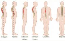
Therefore, it is obvious that the location of the pelvis greatly affects the posture. This is the same as if the central block in the tower is displaced, in which case all blocks above the displacement are at risk of falling. And if you compare the central unit with a box, then the tilt can lead to the box falling out. Similar mechanisms take place when the pelvis is tilted, and the contents of the pelvis are shifted forward. As a result, there is a protruding abdomen and bulging of the buttocks. Since the pelvis is the junction of the upper and lower torso, it plays a key role in body movement and balance. The pelvic bones support the most important supporting part of the body - the spine. In addition, the pelvis allows the lower limbs and torso to move in a coordinated manner (in tandem). When the pelvis is in a normal position, various movements are possible, twisting, tilting and movement biomechanics are balanced and the distribution of load vectors is even. Displacement (skew) of the pelvis from normal positions causes dysfunctional disorders of the spine, as there is a change in the axis of distribution of loads during movement. For example, if there is an axle shift in a car, then the wheels wear out quickly. Something similar happens in the spine, there are effects of leverage and excessive load on certain points, which lead to rapid wear of the structures of the spine. Therefore, often the main cause of pain in the back and neck is a change in the position of the pelvis (displacement, distortion). A change in position changes biomechanics, which can lead to degenerative changes in the spine, disc herniation, scoliosis, osteoarthritis, spinal canal stenosis, sciatica, etc. Pelvic tilt also leads to pain and dysfunction in the neck, neck pain radiating to the shoulders, arms, contributes to the development of carpal tunnel syndrome and other problems in the limbs.
Causes of skew (displacement) of the pelvis
In the first place, pelvic tilt is caused by a simple muscle imbalance. Technology is developing very quickly and a sedentary lifestyle is one of the main reasons for the development of imbalance, because our body requires a certain amount of movement that it does not receive. Prolonged sitting and low physical activity are sufficient conditions for the development of muscle imbalance, leading to a pelvic tilt and, as a result, the appearance of dysfunctional disorders in the spine and the occurrence of back pain.
Accidents and injuries are common causes of pelvic tilt, such as side impact, heavy lifting while twisting, falling to one side, side-carrying, such as carrying a child on your hip or carrying a heavy bag all the time on one shoulder. In women, the pelvis is less stable from birth than in men, since a certain flexibility and elasticity of the pelvic structures is necessary for the normal course of pregnancy and childbirth. Therefore, pregnancy is often the main cause of pelvic displacement in women.
Damage to the pelvic muscles is the most common cause of misalignment. Injured muscles usually harden and shift in order to protect the surrounding structures. If the muscles in the pelvic region, such as the sacrum, are damaged, then the tightening of the muscles will lead to an effect on the ligaments attached to the pelvis and joints. As a result, structures such as the sacroiliac joints will also have a certain disposition. Muscle compaction after injury persists until full muscle recovery and during this period of time the pelvis remains in an abnormal position.
The difference in leg length can also be the cause of the pelvic tilt, and in such cases, the tilt can be from right to left or vice versa. But the displacement can also be forward or backward or it can be twisting of the pelvis.
Many conditions can lead to muscle spasms that cause the pelvis to twist. A herniated disc can cause muscle spasm adaptive nature and, in turn, in antalgic scoliosis with functional pelvic tilt. Active people often experience tension in the calf muscles, which in turn creates tension around the pelvis. Surgery such as hip replacement can also cause the pelvis to reposition itself.
Since the pelvis is one of the most stressed areas of the body due to movement and weight support, movements that cause pain and stiffness are a clear indicator of a pelvic alignment problem. Back pain in particular is a common indicator of pelvic tilt. In addition to participation in the movement in the pelvic cavity are: part of the digestive organs, nerves, blood vessels, reproductive organs. Therefore, in addition to back pain, there may be other symptoms, such as numbness, tingling, bladder and bowel problems, or reproductive problems. Most often, changes in the following muscles lead to pelvic disposition:
M. Psoas major (lumbar muscle) anatomically can lead to extension and flexion of the hip, which leads to an anterior displacement of the pelvis.
M.Quadriceps (quadriceps), especially the rectus muscle, can lead to hip flexion.
M.Lumbar erectors can cause lumbar extension.
M. Guadratus lumborum with bilateral compaction can cause an increase in lumbar extension.
M.Hip adductors (adductors of the thigh) can cause the pelvis to tilt forward as a result of internal rotation of the hip. This leads to shortening of the adductor muscles.
M. Gluteus maximus (gluteus maximus) is responsible for hip extension and is an antagonist of the psoas major muscle.
M.Hamstrings The muscle of the back of the thigh, this muscle can be hardened. The muscle can be weak at the same time hardened due to the fact that it is a synergist of the gluteus maximus muscle and this may be of a compensatory nature. The deep muscles of the abdominal wall, including the transversus abdominis and internal obliques, may tighten due to weakening of the lumbar erectors muscles.
Symptoms
Symptoms of displacement (skew) of the pelvis can be both moderate and severe and significantly impair the functionality of the body. With moderate misalignment, a person may feel unsteady when walking or frequent falls are possible.
The most common symptoms such as pain:
If the pelvis is displaced for a long time, then the body will correct and compensate for the violation of biomechanics and asymmetry and there will be a corresponding adaptation of the muscles, tendons and ligaments. Therefore, treatment may take some time. In addition, pelvic tilt can be difficult to correct, as a pathological stereotype of movements is formed over time. The longer the period of pelvic tilt, the longer it takes to restore normal muscle balance.
Diagnosis and treatment
Pelvic tilt is usually well diagnosed on physical examination of the patient. If it is necessary to diagnose changes in the spine or hip joints instrumental examination methods are prescribed, such as radiography or MRI (CT).
Exist various options treatment of pelvic tilt and these methods depend on the cause that led to the pelvic tilt. In the treatment of, for example, twisting of the pelvis, it is necessary to reduce muscle damage. For this, various methods of physiotherapy, NSAIDs can be used. If the skew of the pelvis is due to the difference in the length of the limbs, then it is necessary to use individual insoles or surgical methods of treatment.
But, in any case, the treatment of pelvic tilt is effective only in combination with the impact on the pathogenetic links that led to a change in the position of the pelvis and a violation of biomechanics (physiotherapy, massage, manual therapy and exercise therapy). Exercise therapy is the leading treatment for pelvic disposition, especially when muscle problems are the cause of the pelvic tilt.
The use of materials is allowed with an active hyperlink to the permanent page of the article.


The influence of the Internet on a person
Types of seizures Tonic-clonic seizures in children
Profession Internet project manager
best remedy for prostatitis
Symptoms and treatment of mastopathy of the mammary glands