MINISTRY OF EDUCATION, SCIENCE AND YOUTH
REPUBLIC OF CRIMEA
CRIMEAN REPUBLICAN NON-SCHOOL EDUCATIONAL INSTITUTION
"CENTER FOR ECOLOGICAL AND NATURALISTIC CREATIVITY
STUDENT YOUTH»
OPEN LABORATORY LESSON:
STUDYING THE STRUCTURE OF THE PLANT CELL
Developed by:
Kuznetsova Elena Yurievna, methodologist of the highest category,
head of the educational team
"Fundamentals of Biology", Ph.D.
Simferopol, 2014
Topic of the lesson: Examining the structure of a plant cell under a microscope
Target: to consolidate and deepen knowledge about the structural features of a plant cell.
Lesson type: lab session
Used forms and methods: conversation, testing, work with microscopic equipment.
Introduced concepts: cell wall, nucleus, vacuole, chlorophyll grains, starch grains, plasmolysis, deplasmolysis.
Materials and equipment: microscopes with accessories, water, 5% saline solution, juicy onion scales, wallisneria leaf, potatoes.
Lesson plan:
Knowledge update. Testing.
The structure of the microscope and work with microscopic equipment.
Method for the manufacture of temporary preparations. Preparation of the preparation of the epidermis of juicy onion scales, microscopy.
Setting up an experiment. The phenomena of plasmolysis and deplasmolysis.
Starch grains of potato pulp.
Chlorophyll grains of Vallisneria leaf.
Lesson progress:
1. Knowledge update. Testing.
Test tasks on the topic "Structure of a plant cell"
1 What organelles are absent in an animal cell:
a) mitochondria b) plastids c) ribosomes d) nucleus
2. In which organelles is primary starch formed:
3. In which organelles does oxidative phosphorylation occur:
a) mitochondria b) chloroplasts c) nucleus d) ribosomes
4. Which group of lipids forms the basis cell membranes:
a) neutral fats b) phospholipids c) waxes d) carotenoids
5. A plant cell, unlike an animal cell, has:
a) endoplasmic reticulum b) Golgi complex
c) vacuole with cell sap d) mitochondria
6. The granular endoplasmic reticulum differs from the agranular one by the presence of:
a) centrosomes b) lysosomes c) ribosomes d) peroxisomes
7. Mitochondria are called the energy stations of the cell. This name of organelles is associated with their function:
a) protein synthesis b) intracellular digestion
c) transport of gases, in particular oxygen d) ATP synthesis
8. The supply of cell nutrients is contained in:
a) nucleus b) chloroplasts c) nucleolus d) leukoplasts
9. In which of these organelles is photophosphorylation carried out:


The structure of the microscope and work with microscopic equipment.

The structure of the mechanical device of the microscope includes a tripod, an object stage, an illumination system, a rack, a micrometric screw, a tube and a revolver.
The object of study is placed on the subject table. A lighting device is located under the subject table; it includes a two-sided mirror. Collecting the rays coming from the light source, the concave mirror reflects them in the form of a beam of rays, which is directed to the object through a hole in the center of the table.
The optical system of a microscope consists of an eyepiece, an objective and a tube connecting them. Lenses are of two kinds: for small and large magnification of the image. If it is necessary to change the lens, they use a revolver - a concave round plate with lenses screwed into it. The entire optical system is mobile: by raising it by rotating the rack counterclockwise or lowering it by rotating it clockwise, they find a position at which the object becomes visible to the observer.
The structure of the microscope:
1 - eyepiece; 2- revolver for changing lenses; 3 - lens;
4 - rack for rough pickup;
5 - micrometer screw for precise aiming; 6 - object table; 7 - mirror; 8 - condenser
3. Methodology for the manufacture of temporary preparations. Preparation of the preparation of the epidermis of juicy onion scales, microscopy.
Prepare a glass slide with a drop of water;
From the fleshy scales of the bulb, cut a small piece (about 1 cm 2) from the inner (concave) side with a scalpel, remove the transparent film (epidermis) with tweezers or a needle. Put in the prepared drop and apply a coverslip;
To study the structure of the cell at low and high magnification;
Draw one cell. Mark the cell wall, the parietal layer of the cytoplasm, the nucleus, the vacuole with cell sap.

The structure of a plant cell
Setting up an experiment. The phenomena of plasmolysis and deplasmolysis.
cook new drug from the skin of an onion. Remove the specimen from the microscope stage, replace the water under the coverslip with 5% common salt (NaCl) solution. The coverslip can be left on: put a drop of the solution near it so that it merges with the water under the glass, and then attach a strip of filter paper on the opposite side. The solution will go under the coverslip and replace the water.
We placed the cell in a hypertonic solution, i.e. the concentration of the solution outside the cell exceeds the concentration of substances in the cell. At the same time, water leaves the vacuole, the volume of the vacuole decreases, the cytoplasm moves away from the membrane and contracts along with the vacuole. There is a phenomenon plasmolysis .
Depending on the degree of concentration of the solution taken, the speed of processing and the shape of the cell, the patterns of plasmolysis may be different.
If plasmolysis proceeds slowly in a weak solution, the contents of the cell most often first move away from the membrane at the ends of the cell (corner plasmolysis), may be affected large plots cells (concave plasmolysis). The contents of the cell can separate into one round drop (convex plasmolysis). When the cell is exposed to a stronger solution, plasmolysis proceeds faster, and there are pictures of convulsive plasmolysis, in which the contents remain connected to the membrane by numerous Hecht threads.

The phenomenon of plasmolysis
A - Plant cell:
1 - cell wall;
2 - vacuole;
3 - parietal layer of the cytoplasm;
4 - core.
B - D - Plasmolysis:
B - corner;
B - concave;
G - convex;
D - convulsive
5 - Hecht threads
During plasmolysis, the cell remains alive. Moreover, an indicator of cell viability can be its ability to plasmolysis. When the cell is returned to clean water comes deplasmolysis , at which the cell absorbs water again, the vacuole increases in volume, and the cytoplasm, pressing against the membrane, stretches it.
sketch different stages plasmolysis with appropriate designations.
Carry out the phenomenon of deplasmolysis by displacing the salt solution from under the coverslip with water and filter paper.
Starch grains of potato pulp
starch grains - the main type of reserve nutrients of a plant cell. They are formed only in plastids of living cells, in their stroma. Grains of assimilation (primary) starch are deposited in chloroplasts in the light, which are formed with an excess of photosynthesis products - sugars.
Prepare a preparation of starch grains from potato pulp. For this purpose, squeeze the juice of the pulp of a potato tuber onto a glass slide into a drop of water. Examine under a microscope, draw.

Starchy potato grains
Vallisneria leaf chlorophyll grains
Prepare a preparation from a Vallisneria leaf, placing rather large cells of the lower third of the leaf blade in the center of the field of view, not far from the midrib. Examine this area under high magnification, sketch the chloroplasts.

Chloroplasts in Vallisneria leaf cells
Lesson conclusions:
Establish the differences between plant and animal cells;
Establish patterns of osmotic phenomena in the cell.
Homework:
Solve the crossword Cell structure»

Crossword "Cell structure"
Horizontally: 2 . Liquid mobile contents of the cell. 5 . The main organelle of the cell. 8 . Component microscope. 10 . unit of a living organism. 12 . A simple magnifying device. 13 . A tube in a microscope with magnifying glasses inserted. 16 . Microscope maker. 18 . Physiological process inherent in a living cell. 19 . On which preparations are prepared. 22 . The area between cells with destroyed intercellular substance, filled with air.
Vertically: 1 . Oculus ( lat.). 3 . Complex optical device. 4 . A thin area in the cell membrane. 6 . The main structure of the nucleus. 7 . Cell cavity filled with cell sap. 9 . The part at the upper end of the microscope tube, consisting of a frame and two magnifying glasses. 11 . The part of the microscope to which the tube is attached. 14 . cell cover. 15 . Small bodies in the cytoplasm of a plant cell. 17 . Part of the bulb from which the drug is prepared. 20 . The part of the microscope located at the lower end of the tube. 21 . An aquatic plant in whose leaf cells one can see the movement of the cytoplasm.
Working process
Investigate preparations obtained from raw and boiled vegetables. To obtain preparations from vegetables, a part of the pulp is separated from each specimen and cut in half. One half is stored in cold water, the other is cooked until tender. To ensure comparability of results, microscopic sections are removed from those places of the pulp that were in contact with each other before cutting before cooking. Soaked bean seeds are divided into two cotyledons, one of which is boiled.
For microscopy, two preparations are placed on each slide: on the left side - from raw products, on the right - from boiled products, adding a drop of water to them. Each preparation is considered in unstained and stained form. As dyes for preparations from vegetables, safranin is used, which colors pectin substances in orange-yellow color, and fiber and flakes of denatured proteins - in cherry-red, in addition, iodine is used for starchy vegetables. Bean preparations are stained only with iodine, which stains starch grains blue-black, and the protein matrix and cell walls golden yellow.
When staining preparations, water is removed from them with filter paper, a drop of paint is applied and incubated for two minutes. Then, the excess of the coloring matter is removed from the preparations and a drop of water is added to them. Cover slips are placed on stained and unstained preparations.
Microscopy of preparations is carried out first at low magnification, and then at high magnification. Draw preparations at high magnification.
1. The study of the structure of the tissues of potatoes and root crops.
From the middle of the peeled tuber (root crop), cut a slice 5 mm thick and cut it in half. Place one half in a glass of cold water, the other half in a glass of boiling water and cook for 10-15 minutes. From the raw and boiled parts of the tuber (root) cut, observing symmetry, one bar with a cross section of 5 × 5 mm. Using a razor blade, make two transparent cuts with an area of 2-4 mm 2 on the end side of each bar. Transfer them with a needle to three glass slides and add a drop of water.
Leave the preparations on one slide unstained, on the other - stain with iodine, on the third - with safranin and iodine. Cover the preparations with glass slides and examine under a microscope. Pay attention to the shape of the cells, their tightness to each other, the state of the cell walls, starch grains in the tissues of raw and boiled potatoes (root crops).
2. Studying the structure of onion tissues. Separate the fleshy scales from the bulb and cut it in half along the growth axis, place one half in a glass of cold water, and boil the other for 15 minutes. With inside of raw and boiled scales, remove a thin film with a dissecting needle. Straighten the resulting films. Cut out from the thinnest sections two preparations with an area of 2 × 2 mm 2 and place them on two glass slides, adding a drop of water to each preparation. Leave the slides unstained on one slide and stain with safranin on the other. Cover prepared preparations with coverslips and examine under a microscope. Pay attention to the thickness and condition of the cell walls, their tightness to each other, the degree of transparency of the contents of the cells, the presence of nuclei. Note the differences in the structure of raw and boiled onion tissues, as well as in the structure and color intensity of individual cell elements.
Use unstained preparations to observe cell plasmolysis. Remove coverslips from preparations, remove water with filter paper and add a few drops of 10% sodium chloride solution, hold for 5-10 minutes, cover with coverslips and examine again under a microscope. Find plasmolyzed cells in the field of view in raw onion preparations, explain the absence of such cells in the boiled onion preparation. Make sketches.
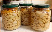
3. The study of the structure of bean seed tissues. Divide the pre-soaked bean seed into two cotyledons, one of which is boiled for 1 hour. From each cotyledon, make two cuts for preparing preparations, unstained and stained with iodine. When examining preparations under a microscope, pay attention to the difference in the structure of the tissues of raw and boiled bean seeds.
Draw conclusions about the effect of thermal cooking on the structure of vegetable tissues.
Task number 2. To study the influence of technological factors on
Preservation of potato cell walls during production
mashed potatoes
Working process
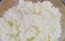
Option 1. The two side parts of the potato tuber remaining from the previous study are placed in a glass of boiling water and boiled for 20-25 minutes. Grind one part in a hot state in a mortar, cool the other to room temperature and also grind.
Prepare preparations for microscopy. On a glass slide, transfer a little of both purees with a dissecting needle, add a drop of iodine solution and cover with coverslips. When considering preparations at low magnification, compare the number of cells with destroyed cell walls in both purees. Examine preparations at high magnification and sketch. Make a conclusion about the effect of the temperature of boiled potatoes when mashed on the degree of preservation of cell walls.
Option 2. Carry out comparative microscopy of dry mashed potatoes and reconstituted by liquid with and without subsequent stirring.
Weigh out two samples of dry puree weighing 25 g and place them in two glasses. In two other glasses, heat up to 78 - 80 ° C, 100 cm 3 of water each and pour dry puree with it. Close one glass with a watch glass and let the puree swell for 2 minutes. Prepare preparations for microscopy from dry puree and reconstituted puree. With the end of a glass rod moistened with water, take some dry puree and place it on a glass slide, add a drop of water, then stain with iodine, cover with a cover slip and examine under a microscope. Note the presence of cells with destroyed cell walls in the dry puree. Prepare preparations from the reconstituted puree and examine them under a microscope, as indicated in option 1.
Compare the number of cells with destroyed cell walls in fresh potato puree, mashed hot, and in dry puree, as well as in reconstituted puree. Draw drugs.
Purpose: To get acquainted with the structure of starch grains of the main food plants
Methodical instructions. The most common storage substance in plants is the polysaccharide starch. Primary starch is formed from the products of photosynthesis in the leaves of plants and has the form of small grains. Here it is not stored, but transported to build plant organs or deposited as a reserve substance in fruits.
Rice. 6. Starch grains various kinds plants
A - from potato tubers: 1 - simple; 2 - complex; 3 - semi-complex;
B - wheat (simple); B - oats (complex); G - corn (simple);
D - rice (complex); E - buckwheat (simple)
Here it is not stored, but transported to build plant organs or deposited as a reserve substance in fruits.
Secondary or reserve starch is formed in leukoplasts (amyloplasts) in specialized organs - rhizomes, tubers, seeds, fruits. From this starch, simple, semi-complex and complex grains are formed.
If there is one point in the leukoplast, around which layers of starch are deposited, then a simple starch grain is formed (Fig. A1, B, D).
A complex grain is formed if there are two or more deposition points (Fig. A2; C, E, F).
Semi-complex grains are formed if starch is first deposited around several points, and then, after their contact, common layers are formed (Fig. 6, A3). Wheat, rye, corn have simple starch grains, while rice, oats, and buckwheat have complex starch grains. All three types of starch grains are found in potato tubers. The shape, size, structure of starch grains are specific for each plant species. Therefore, when analyzing food raw materials of plant origin, in particular flour, it is possible to identify and establish the presence of impurities in them by the structure of starch grains.
Exercise: Prepare preparations of starch grains of potatoes, wheat, oats, rice, buckwheat. Stain (react) with iodine solution. Draw at high magnification the starch grains of the above plants, while maintaining the proportions between them. Sign the drawings, indicating the type of plant and the type of starch grains.
Work sequence:
Starchy potato grains. A small piece of the tuber is cut off and a smear is made on a glass slide with a drop of water previously applied to it. The drop is covered with a cover glass, microscoped at low, then at high magnification. It is necessary to try to find all three types of starch grains (sometimes this cannot be done). When considering the layering of starch grains, cover the diaphragm and slightly rotate the microscrew. Draw the picture you see.
The preparation is stained with an iodine solution and, looking through a microscope, the staining process is observed.
Preparations of starch grains of wheat, oats, rice and buckwheat are best prepared from swollen seeds. At the same time, cutting the caryopsis, extract its contents (endosperm) and transfer it to a drop of water on a glass slide. Then proceed as in the previous case, and consider at high magnification.
It is necessary to sketch the shape of starch grains of wheat, oats, rice and buckwheat. It is necessary to learn how to differentiate them by structure and determine the species.
Tubers differ from rhizomes in that their stem is short and thick, and the leaves are underdeveloped. As with any shoot, they have buds and are located at the top and in the axils of underdeveloped leaves. Adventitious roots do not develop on tubers. Potato tubers do not grow immediately from underground buds. First, a long white underground shoot grows from the kidney - a stolon. Stolon lives less than a year. The top begins to thicken over time and turns into a tuber by autumn.
A lot of starch accumulates in the tuber in the form of small grains. A potato tuber is a modified shoot with a thickened stem and small leaves.
What to do. Consider the external structure of a potato tuber.
What to watch. Find on its surface the apical and axillary buds (eyes), scars from leaves (brows) and a scar from a separated stolon.
What to do. Count the number of eyes on the tuber.
What to watch. Locate the top and bottom of the tuber.

Note the uneven distribution of eyes on the thickened stem.
That part of the tuber, where there are more eyes, is called the top, and the opposite, where the scar from the stolon is called the base.
What to do. Cut the tuber into two pieces. Drop a drop of iodine solution on the cut of the tuber.
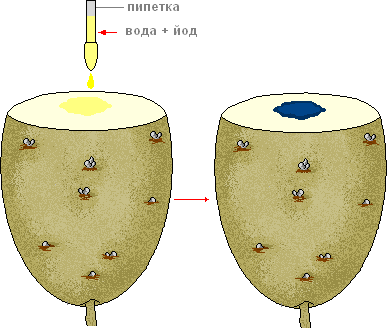
- How did the color of the tuber section change?
- What substances are deposited in the tuber cells?
- What is the importance of a tuber in the life of a plant?
Prepare for a report. Draw in a notebook appearance tuber and label its parts. Write down the signs that prove that the tuber is a shoot.
The tissue (pulp) of potatoes, vegetables and fruits consists of thin-walled cells that grow approximately equally in all directions. This tissue is called parenchyma. The contents of individual cells is a semi-liquid mass - the cytoplasm, into which various cellular elements (organelles) are immersed - vacuoles, plastids, nuclei, starch grains, etc. (Fig. 9.2). All cell organelles are surrounded by membranes. Each cell is covered with a shell, which is the primary cell wall.
The shells of each two neighboring cells are fastened with the help of the middle plates, forming the backbone of the parenchymal tissue (Fig. 9.3).
Contact between the contents of the cells is carried out through plasmodesmata, which are thin cytoplasmic strands passing through the membranes.
The surface of individual specimens of vegetables and fruits is covered with an integumentary tissue - epidermis (fruits, ground vegetables) or periderm (potatoes, beets, turnips, etc.).
Because in fresh vegetables contains a significant amount of water, then all the structural elements of their parenchymal tissue are hydrated to one degree or another. Water as a solvent has an important effect on the mechanical properties of plant tissue. By hydrating to some extent hydrophilic compounds, it plasticizes the structure of the walls and middle plates. This provides a sufficiently high turgor pressure in the tissues.
Turgor is a state of tension arising from the pressure of the contents of the cells on their elastic membranes and the pressure of the membranes on the contents of the cells.
Turgor pressure can decrease, for example, when vegetables and fruits wither or dry out, or increase, which is observed when wilted vegetables are immersed in water. This property of vegetables and fruits can be taken into account in their culinary processing. So, potatoes and root crops with a weakened turgor are recommended to be soaked for several hours before mechanical cleaning to reduce processing time and reduce the amount of waste.
Rice. 9.2. The structure of a plant cell
Rice. 9.3. Plant tissue wall:
1 -- middle plate; 2 - plasmalemma.
Magnification x 45000 (according to J.-C. Roland, A. Seleshi, D. Seleshi)

The vacuole is the largest element located in the center of the cell. It is a kind of bubble filled with cell sap, and is the most hydrated element of the vegetable and fruit parenchyma cell (95 ... 98% water). The composition of the dry residue of cell sap includes, in one amount or another, almost all water-soluble nutrients.
The main mass of sugars contained in potatoes, vegetables and fruits in a free state, soluble pectin, organic acids, water-soluble vitamins and polyphenolic compounds is concentrated in vacuoles.
The cell sap contains approximately 60 ... 80% of minerals from their total amount in vegetables and fruits. Salts of monovalent metals (potassium, sodium, etc.) are almost completely concentrated in the cell sap. Salts of calcium, iron, copper, magnesium are contained in it somewhat less, since they are part of other tissue elements.
Cell sap contains both free amino acids and soluble proteins, which form solutions of relatively low concentration in vacuoles.
A thin layer of cytoplasm with other organelles occupies a near-wall position in the cell. The cytoplasm consists mainly of proteins, enzymes and a small amount of lipids (the ratio of proteins and lipids is 90:1). In the cytoplasm, as in vacuoles, they are in the form of a solution, but more concentrated (10%).
Plastids are organelles that are found only in plant cells. The most typical of these are chloroplasts, which contain chlorophyll. Under certain physiological conditions, plastids do not form chlorophyll; in these cases, they produce either proteins (proteoplasts) or lipids and pigments (chromoplasts), but most often such plastids perform reserve functions, and then starch (amyloplasts) accumulates in them, so plastids are colored and colorless. The latter are called leukoplasts.
The composition of chloroplasts, in addition to chlorophyll, includes proteins and lipids in a ratio of 40:30, as well as starch grains.
During the development of chromoplasts, large globules or crystals containing carotenoids, including carotenes, are formed. The presence of these pigments in green vegetables and some fruits (gooseberries, grapes, renklod plums, etc.) causes different shades of their green-yellow color. Carotenes give a yellow-orange color to carrots, turnips, etc. However, orange color does not always indicate their high content in fruits and vegetables; for example, the color of oranges, tangerines is due to another pigment - cryptoxanthin. At the same time, the relatively high content of carotene in green vegetables can be masked by chlorophyll.
Amyloplasts are filled mainly with large starch granules. It should be noted that in plant cells, all the starch grains contained in them are located in a space limited by the shell of amyloplasts or other plastids.
The cell nucleus contains chromatin (despiralized chromosomes), consisting of DNA and basic proteins (histones), and nucleoli rich in RNA.
Membranes are an active molecular complex capable of exchanging substances and energy.
The cytoplasm at the border with the cell wall is covered with a simple membrane called the plasmalemma. The outer edge of the plasmalemma can be seen by examining under a microscope plant tissue preparations treated with a concentrated saline solution. Due to the difference between the osmotic pressure inside the cell and outside it, water moves from the cell to environment, causing plasmolysis - separation of the cytoplasm from the cell membrane. Similarly, plasmolysis can be induced by treating sections of plant tissue with concentrated solutions of sugars or acids.
Cytoplasmic membranes regulate cell permeability by selectively retaining or passing molecules and ions of certain substances into and out of the cell.
The vacuole, like the cytoplasm, is also surrounded by a simple membrane called the tonoplast.
The main structural components of membranes are proteins and polar lipids (phospholipids). Exist Various types structures of the cytoplasmic membrane: three-layer (from two layers of protein with a biomolecular layer of lipids), granular (from particles whose diameter is about 100 10-10 m, or from smaller particles - subunits). At present, the membrane is considered as a liquid structure penetrated by proteins.
The surface of nuclei, plastids and other cytoplasmic structures is covered with a double membrane consisting of two rows of simple membranes separated by a perinuclear space. These membranes also prevent mixing of the contents of two neighboring organelles. Individual substances pass from one organelle to another only in strictly defined quantities necessary for the flow of physiological processes in tissues.
Cell walls in combination with the middle plates are called cell walls. Unlike membranes, they are characterized by complete permeability.
Cell walls make up 0.7 ... 5.0% of the fresh weight of vegetables and fruits. So, in vegetables of the fruit group, for example, in zucchini, their number does not exceed 0.7%. AT leafy vegetables - white cabbage, lettuce, spinach - about 2%. Root crops differ in the highest content of cell walls - 2 ... 4%.
Cell walls are mainly composed of polysaccharides (80...95%) - cellulose, hemicelluloses and protopectin, therefore they are often called cell wall carbohydrates. The composition of the cell membranes includes all of the above polysaccharides. It is believed that the middle plates consist mainly of acidic polysaccharides (protopectin), which play the role of an intercellular cementing substance, which is sometimes accompanied by protein compounds, and in the oldest tissues - lignin.
Tab.9.1. The content of extensin and hydroxyproline
in the cell walls of some plant foods(%)
In addition to carbohydrates, the cell walls contain nitrogenous substances, lignin, lipids, waxes, and minerals.
Of the nitrogenous substances in the cell walls of plant tissue, a structural protein of extension was found - a polymer from the group of glycoproteins, the protein part of which is associated with carbohydrates - the residues of arabinose and galactose. The molecular weight of the protein part of such macromolecules is 50,000, the extension has the form of a rigid rod, 50% consists of hydroxyproline. The cell wall contains several protein fractions that differ in the content of hydroxyproline.
Extensions in some respects resemble the protein collagen, which performs similar functions in animal tissues. The content of extensin and hydroxyproline in the cell walls of various vegetables and potatoes is not the same (Table 9.1). The cell walls of a potato consist of about 1/5 of extensin. In the cell walls of root crops, it is contained 2 times less than in the cell walls of potatoes; in the cell walls of melon, the content of extensin does not exceed 5%.
The ratio of carbohydrates and extensin in cell walls depends on the type of plant tissue. The cell walls of many plant foods are about 1/3 cellulose, 1/3 hemicellulose, and 1/3 pectin and protein. In the cell walls of tomatoes, there is another 1:1 ratio between carbohydrates and protein.
Lignin is a natural polymer of complex structure that forms the cell walls of plants. It plays the role of an encrusting substance that holds together cellulose and hemicellulose fibers. It is covalently bound to hemicellulose polysaccharides (xplan), pectins and protein. The content of lignin in plant tissues depends on their type and degree of lignification. A significant amount of lignin is contained in the cell walls of beets, carrots, less accumulates in white cabbage.
Due to the fact that the softening of potatoes, vegetables and fruits, which occurs during their thermal cooking, is associated with the destruction of cell walls, it seems appropriate to consider the structure of the latter.
According to modern concepts, the cell wall is a highly specialized aggregate consisting of various polymers (cellulose, hemicellulose, pectin, proteins, etc.), the structure of which in different plants is encoded with the same degree of accuracy as the structure of protein molecules.
On fig. 9.4 shows a model of the structure of the primary cell wall.
The primary cell wall consists of fibers (microfibrils) of cellulose, which occupy less than 20% of the volume of the hydrated wall. Being parallel in the cell walls, cellulose fibers form micelles with the help of hydrogen bonds, which have a regular, almost crystalline packing. One cellulose micelle can be separated from another by a distance equal to ten of its diameters. The space between cellulose micelles is filled with an amorphous basic substance (matrix) consisting of pectin substances, hemicelluloses (xyloglucan and arbinogalantan) and a structural protein associated with tetrasaccharides.
The primary cell wall is considered as a whole bag-like macromolecule, the components of which are closely interconnected. Numerous hydrogen bonds exist between cellulose micelles and xyloglucan. In turn, xyloglucan is covalently linked to the galactan side chains of pectin substances, and pectin substances through arabinogalactan are covalently linked to the structural protein.
Considering that the cell walls of many vegetables and fruits are characterized by a relatively high content of divalent cations, mainly Ca and Mg (0.5 ... 1.0%), chelate bonds in the form of salt bridges.
Rice. 9.4. The structure of the primary cell wall (according to Albersheim):
1 - cellulose microfibril: 2 - xyloglucan; 3 - main
rhamnogalacturonic chains of pectin substances; 4 - side
galactan chains of pectin substances; 5-structural protein
with arabinose tetrasaccharides; 6- arabinogalactan

The probability of formation of salt bridges and the degree of esterification of polygalacturonic acids are inversely related. Salt bridges contribute to the strengthening of cell walls and parenchymal tissue in general.
The integumentary tissues of potato tubers, root crops and other vegetables are characterized by a reduced nutritional value due to the concentration of fiber and hemicelluloses in them, therefore, during the cooking of potatoes and most vegetables, these tissues are removed.


Sun skirt: types and how to wear it Black sun skirt with a T-shirt how to wear
Ground bird cherry Ground bird cherry cook
Own business: production of chips
How to care for your skin in spring Face masks in spring
Seizures in the corners of the mouth: causes and treatment in adults and children