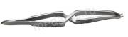
Surgical Instruments haemostatic - clamping instruments are used to grasp and clamp tissues and organs in a wound in order to temporarily stop bleeding, block the lumen of hollow organs, to stop the flow of liquid content in them, crush walls, grab and strengthen surgical drapes, drainage tubes, etc. According to the instructions, clamping surgical instruments are divided into several types: locking, hinged, spring and screw. To reduce the sliding of the tool, notches or grooves are usually made on its working parts longitudinal and transverse to the tool axis. The shape of the clamping tools can be straight and curved (axial, plane). This group includes hemostatic clamps the following types: Kocher (toothed), Billroth (grooved), Halstead straight and curved - "mosquitoes".
Buy hemostatic surgical instruments
Surgical clamp
Hemostatic clamp
Intestinal clamp
Kornzang surgical
Surgical forceps
Surgical forceps
Language holder
Surgical clamps
Clip Alice for gripping the intestinal wall Art. Z-24 P
Clamp for grasping the intestinal wall
The working parts of the intestinal clamps are elastic metal plates, the width of the working part is 6 mm
length 152 mm
Number of teeth 4x5
Pakistan
Price: RUB 408.00
Art. K-132 P
 Clips for fixing surgical drapes are intended for:
- to delimit the operating field from the rest of the skin surface;
- to delimit the opened cavities (abdominal, chest) from the edges of the wound.
Requirements for clamps for surgical drapes:
- strength;
- versatility for fastening the edges of linen of various thicknesses;
- exclusion of self-opening of working parts;
- reliability of fixing the linen.
The clamps for fixing the operating linen do not slide off the linen under the influence of a load of up to 10 kg.
Length: 90mm
Manufacturer: Surgicon Pvt LTD, Pakistan
Price: RUB329.00
Hemostatic clamp
Hemostatic clamp: the general name for surgical clamps for clamping blood vessels to temporarily stop bleeding; hemostatic clamps have working jaws with a fine notch and a conical outer surface.
Hemostatic forceps "Mosquito"
 Clamp type "Mosquito" also called the Halstead clamp. Hemostatic clamp "Mosquito" has the thinnest working surfaces.
curved for newborns apply dfor hemostasis of small vessels in neurosurgical operations and in pediatric practice.
Manufacturer: Surgicon Pvt LTD, Pakistan
| vendor code | Name | Length | length of the working part | Price, RUB |
| Z-62-1 P | Hemostatic clamp "Mosquito" straight | 160 mm | 211,00 | |
| Z-62-2 P | Hemostatic clamp "Mosquito" curved | |||
| Z-62-4 P | Hemostatic clamp "Mosquito" curved along the edge | 154 mm | 190,00 | |
| Z-120 P | Hemostatic clamp "Mosquito" straight for newborns | 125 mm | 20 mm | 182,00 |
| Z- 121 P | Hemostatic clamp "Mosquito" curved for newborns | |||
| Z- 122 P | Hemostatic clamp "Mosquito" curved along the edge for newborns |
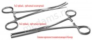 with long narrow working jaws with sharp teeth, and the only tooth of one jaw enters between two teeth of the second jaw, have teeth on the gripping surfaces, which injures the tissues, but captures them firmly.
Manufacturer: Surgicon Pvt LTD, Pakistan
| vendor code | Name | length | length of the working part | Price in rub. |
| Z-5 P | Hemostatic clamp 1x2 tooth, serrated, straight No. 2 | 160 mm | 83 mm | 201,00 |
| Z-5-1 P | Hemostatic forceps 1x2 tooth, serrated, curved No. 2 | 160 mm | - | 220,00 |
| Z-21 P | Hemostatic clamp 1x2 tooth, serrated, straight No. 1 | 150 mm | 34 mm | 190,00 |
| Z-21-1 P | Hemostatic forceps 1x2 tooth, serrated, curved No. 1 | 150 mm | 25 mm | |
| Z-3 1 P | Hemostatic clamp 1x2 tooth, serrated, straight No. 3 | 200 mm | 25 mm | 262,00 |
| Z-31-1 P | Hemostatic forceps 1x2 tooth, serrated, curved No. 3 | 200 mm | 60 mm |
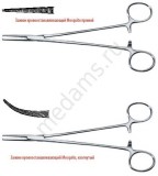
Hemostatic forceps Billroth
 Clamp hemostatic Billroth designed for gripping and clamping vessels. Has working jaws with a fine notch and a conical outer surface. Notches on the gripping branches injure tissue less, but not firmly capture them.
Manufacturer: Surgicon Pvt LTD, Pakistan
| vendor code | Name | length | length of the working part | Price in rubles |
| Z-92 P | Hemostatic serrated clamp, straight No. 1 | 160 mm | - | 201,00 |
| Z-53 P | Hemostatic serrated clamp, curved No. 1 | 158 mm | 40 mm | |
| Z-32 P | Hemostatic serrated clamp, straight No. 2 | 198 mm | - | 262,00 |
| Z-37 P | Hemostatic serrated clamp, curved No. 2 | 196 mm | 25 mm | 270,00 |
| Z-42 P | Hemostatic serrated clamp, curved No. 3 | 270 mm | - | 301,00 |
| Z-42-1 P | Hemostatic serrated clamp, straight No. 3 | 270 mm | - | |
| Z-3 P | Hemostatic forceps serrated, vertically curved | 240 mm | - | 663,00 |
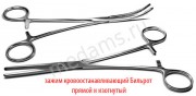
Hemostatic forceps with atraumatic cutting, straight and curved
Hemostatic clamp with atraumatic cutting for grasping and clamping blood vessels, the general name of surgical clamps for clamping blood vessels in order to temporarily stop bleeding; hemostatic clamps have working jaws with a fine notch and a conical outer surface.
Manufacturer: Surgicon Pvt LTD, Pakistan
Popper Clamp is a long straight surgical forceps used in gallbladder surgery.
Manufacturer: Surgicon Pvt LTD, Pakistan
Intestinal elastic clamp
Intestinal clamps applied to hollow organs (stomach, small intestine and large intestine) to achieve the following goals:
- delimitation of damaged areas;
- performance of high-quality linear cuts of the wall;
- separation of the operating field from the infected organ contents;
- overlapping the lumen of the organ.
Manufacturer: Surgicon Pvt LTD, Pakistan
| vendor code | Name | Length | Working part length | Price in rub. |
| Z-40-1t P | Elastic intestinal forceps for adults, straight | 240 mm | - | 369,00 |
| Z-40-2t P | Elastic intestinal forceps for adults, curved | 235 mm | - | 441,00 |
| Z-40-3t P | 200 mm | - | 567,60 | |
| Z-40-4t P | 192 mm | - | 423,70 | |
| Z-40-5t P | Elastic intestinal forceps for children, straight | 170 mm | - | 274,60 |
| Z-40-6t P | Elastic intestinal forceps for children, curved | 161 mm | - | 314,20 |
The hemostatic clamp is designed to clamp blood vessels in deep cavities.
Manufacturer: Surgicon Pvt LTD, Pakistan
 Clamp Hook- designed for attaching surgical drapes to the skin.
Manufacturer: Surgicon Pvt LTD, Pakistan
Kornzang
 Kornzang a surgical instrument for grasping and supplying sterile instruments and dressings, which is a clamp with a ratchet, long straight or curved jaws and oval notched jaws Made of durable and high-quality medical steel that can withstand multiple sterilizations.
Manufacturer: Surgicon Pvt LTD, Pakistan
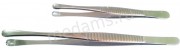
Tweezers
Tweezers- serve to grip and hold various tissues. Each tweezer consists of two steel plates (jaws) with spring properties. Some ends of the plates are soldered (or welded) together. Opposite ends, diverging wedge-shaped, are called paws or jaws. On the outer side of the plates, in the middle part, there are support plates for fingers, which have longitudinal small corrugations or notches. Tweezers quality: When closing the tweezers, the teeth or notches on one sponge should fit snugly into the recesses or notches on the other. The strength of the sponges is checked by compressing a rubber tube with a diameter of 4-6 mm, and for small tweezers with a diameter of 4 mm. There should be no skewing when closing the jaws. Free convergence is checked by clamping the writing paper (the imprint of the sponges must be preserved).
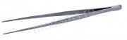
 Forceps anatomical- has transverse notches on the working surface, which makes it possible to hold fabrics more reliably. Tweezers length 150, 200 and 250 mm with different jaw widths.
 Surgical forceps on the working surface of the ends of the jaws of this tweezers there are teeth: on one jaw one, on the other - two, between which, when the tweezers are closed, the tooth of the first jaw fits tightly. Tweezers length 150, 200 and 250 mm with different jaw widths.
 - a surgical instrument used to apply staples when suturing a wound.
Vascular forceps it is used in surgery when manipulating large and medium-sized vessels, allowing them to be held without trauma.
 Coagulation forceps- a surgical instrument designed for coagulation of soft tissues and blood vessels.
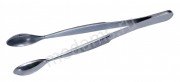 Tweezers spoon intended for the distribution of tablets and powders in medical and health institutions.
Tweezers Brown Adson- Classic tweezers, wide areas for the surgeon's fingers make it easy to hold tissues. 8x9 in-line teeth provide a secure fit. 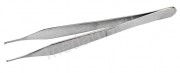 - Surgical tweezers are designed for grasping biological tissues and dressings.
 designed for grasping dense tissues (tendons, etc.), as well as needles, ligatures, etc. The working surface of the jaws of this tweezers is presented in the form of paws, along the edges of which teeth are located. The gripping power of this tweezers is significantly greater than that of others. Available in lengths of 150 and 200 mm.
Manufacturer: Surgicon Pvt LTD, Pakistan
| vendor code | Name | Dimensions (edit) | Price in rub. |
| P-14 | 369,00 | ||
| PM-26 P | 150x1.5 mm | 923,20 | |
| PM-29 P | Vascular forceps with atraumatic cutting | 200x1.5 mm | |
| PM-30 P | Vascular forceps with atraumatic cutting | 250x1.5 mm | 339,60 |
| P-40 P | Coagulation forceps | 200x1.0 mm | 214,80 |
| P-22 P | Tweezers-spoon for dispensing tablets and powders | 140x17 mm | 255,00 |
| PM-40 | Tweezers "Brown-Adson" fabric | 121 mm | 298,80 |
| PM-42 | Adson's tweezers (rat tooth) | 121 mm | 376,50 |
| PM-42-1 | Adson's tweezers (rat tooth) | 150 mm | |
| PM-11 P | Forceps anatomical | 150 mm | 79,00 |
| PM-12 P | Forceps anatomical | 200 mm | 109,00 |
| PM-17 P | Forceps anatomical | 250 mm | 132,00 |
| PM-8 P | Surgical forceps | 150 mm | 85,00 |
| PM-9 P | Surgical forceps | 200 mm | 120,00 |
| PM-10 P | Surgical forceps | 250 mm | 145,00 |
| P-83 P | Toothed tweezers | 150 mm | 249,00 |
| P-157 P | Toothed tweezers | 200 mm | 397,00 |
Forceps
Surgical forceps used in surgical operations involving hollow organs and soft tissues.
In AMS-Med, you can buy the following types of surgical forceps:
- tool pliers straight or curved - designed to squeeze or grip any tools;
- bayonet pliers with narrow oval jaws - used in various surgical procedures for gripping
and retention;
- bone forceps- forceps with semicircular notches on the cheeks, designed to hold bone fragments during osteosynthesis;
- bone nippers- used for biting bone fragments - when treating wounds of the brain and facial parts of the head.
Luer Nippers They are distinguished by the rounded shape of the working part with a cavity inside, into which the bitten off bone fragment is placed.
Liston pliers are made as side cutters and provide a relatively thin and straight cutting line. To increase the cutting torque, a double gear is installed in the nippers.
Dahlgren's Nippers differ in that the cutting part is made in the form of a hook and it can be replaced in case of breakage or wear. These forceps are used for trepanning of the bones of the cranial vault;
- hemorrhoidal forceps- forceps with holes and annular grooves at the ends of the jaws, designed to grasp and hold hemorrhoids during surgery;
- sequestral forceps- forceps with straight or curved jaws, having a slanting cut, designed to remove bone sequesters and bone fragments.
Manufacturer: Surgicon Pvt LTD, Pakistan
| vendor code | Name | Dimensions (edit) | Price in rub. |
| Shch-34t | Pick-up pliers, straight | 280 mm | 434,30 |
| Shch-35 | Pick-up pliers, curved | 280 mm | 526,70 |
| Sch-45 P | Bayonet pliers with narrow oval jaws | 1605,50 | |
| Sch-98 P | Bone nippers articulated with double gear with straight jaws, curved along the plane | 230 mm | 2926,00 |
| Shch-113 P | Bone forceps, articulated nippers with double gear with narrow oval jaws, curved along the plane | 180 mm | 3047,70 |
| Shch-59 P | Bone nippers with round jaws, straight | 170 mm | 1511,50 |
| Shch-61 P | Bone nippers with round jaws, curved | 170 mm | 1732,50 |
| Shch-55-1 P | Hemorrhoidal forceps, fenestrated, curved | 215 mm | 502,00 |
| Shch-55-2 P | Hemorrhoidal forceps, fenestrated, straight | 215 mm | 435,10 |
| Sch-107 P | Sequestral forceps, curved No. 1 | 1262,80 | |
| Sch-102 P | Sequestral forceps, curved No. 2 |
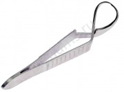
Language holders
Language holder is a tool for grasping, pulling and holding the tongue during surgical operations, it is a clamp with a rack and sponges of various shapes... Language holders for children and adults are presented.
Manufacturer: Surgicon Pvt LTD, Pakistan
SURGICAL CLAMPS- medical instruments designed for clamping organs, tissues and objects during surgery.
3.x. according to their functional purpose, they are divided into clamps, holders and tongs. With the help of clamps, the lumen of hollow organs is closed to stop the movement of contents in them, clamping of blood vessels to temporarily stop bleeding or blood flow in them. Holders are used to hold organs, tissues, materials and objects in a certain position. With forceps they grab and move (pull, remove) organs, tissues and material (see. Medical forceps).
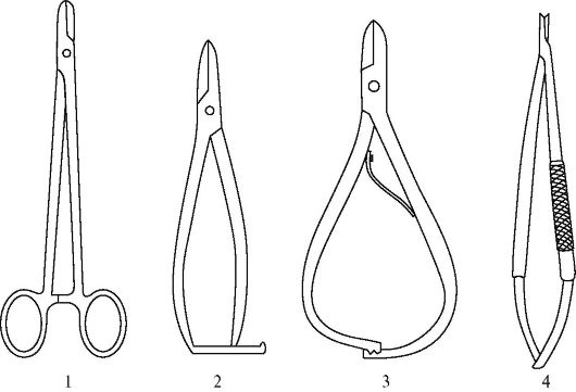
3.x. have, as a rule, two working jaws, between which an organ, tissue, material or object is clamped. Depending on the method of converging and closing the working parts of the tool, they are divided into the following types: spring (clamp, tweezers), hinged, slide and screw (Fig. 1). In addition, there are combined clamps, for example, hinged-slide or spring-screw. The most important functional characteristic clamping tool is the amount of pressure exerted by working jaws on organs or tissues placed between them. The amount of this pressure per unit area of tissue clamped in the clamp sometimes needs to be adjusted so that it does not exceed a certain value in order to avoid injury. Such rationing is carried out, for example, in clamps intended for temporary clamping of hollow organs. A clamping tool that does not cause changes in the structure of tissues is called elastic, and those that cause some reversible changes in tissues are called rigid. There are also crush clamping tools that crush tissue.
3.x. have been known for a long time. Many well-known surgeons took part in their creation and improvement (some types of 3.x. Retained their names in their names). Continuous improvement 3.x., Associated with the development of surgical techniques and the emergence of new materials and technologies. techniques used in their manufacture, has led to the fact that modern clamps differ significantly in outward appearance from those originally suggested by the authors. 3.x. are made of high quality stainless steel, their outer surfaces must be polished, the inner surfaces (teeth, notches, bunny) can be matte.
Features 3.x. are determined by a number of quantitative indicators that can be monitored using instruments. So, for example, when using 3.x. the effort is known, a cut must be applied by the surgeon when working with the tool, and the effort that develops on the working parts 3. x. This goes a long way in making the 3.x easier to work with and provide the necessary compression. fabrics and objects. Curvatures and bends 3.x. are determined depending on the conditions of their use. Most often 3.x. - these are hinged-type clamps with a ratchet.
Clamps are subdivided into hemostatic and hollow clamps.
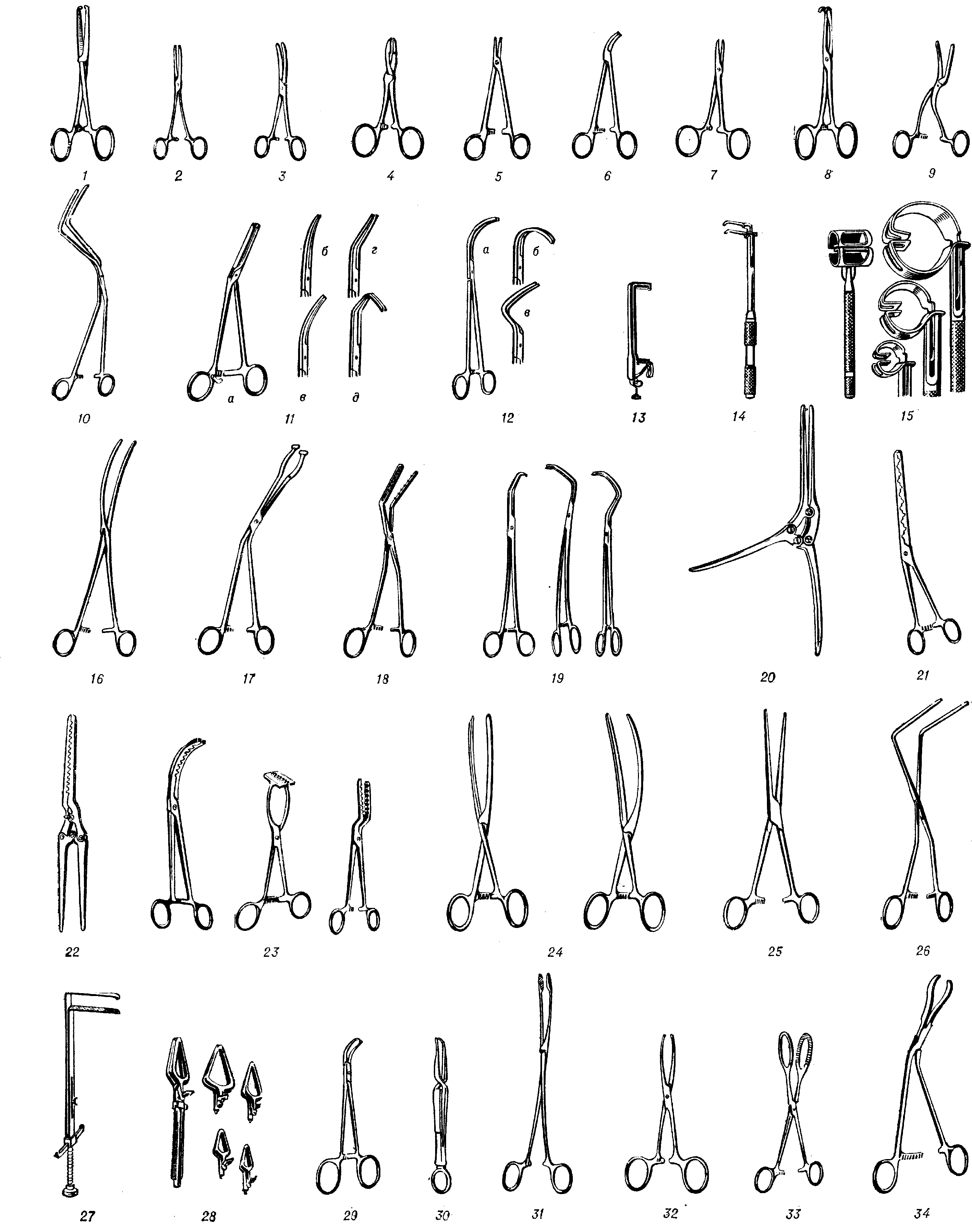
Hemostatic clamps are designed to temporarily stop bleeding from the lumen of a transected vessel. They are used in all areas of surgery. They have a box lock and a rectangular handle. In general surgery, the most widely used are: Kocher's clamp - a straight clamp with cutting on the working jaws and sharp teeth at the ends (Fig. 2, 1) and without them (Fig. 2, 2) 15, 16, 20 and 22 cm long; Billroth's clamp is curved, 16 cm long (Fig. 2.3) and a clamp with Pean's oval jaws (Fig. 2.4). In neurosurgery, straight clamps are used (Fig. 2.5), curved (Fig. 2, 6) 14.5 cm long with an oblique notch on narrow jaws and Mosquito-type clamps (Fig. 2.7) - the most elastic of the hemostatic clamps with a length 7.5 and 15 cm, which are produced straight and curved along the plane and along the edge. In ophthalmology, hemostatic clamps with a length of 8.5 cm, elastic with small teeth, are used and the clamping force on the third tooth of the ratchet is only 0.7 kgf. For surgical interventions in deep cavities, straight and curved clamps are used. They are distinguished by the sharply bent ends of the working jaws (Fig. 2.8) and sometimes by the presence on the jaws, in addition to the transverse notch, of grooves directed along the working jaw.
The outer surfaces of the working sponges, hemostatic clamps should be made so that the ligature, which is previously tied on them, easily slides from the clamp onto the vessel. Therefore, the jaws of these clamps are made conical, and the cleanliness of their surface must be at least 10th class in accordance with GOST. For short-term clamping of blood vessels in order to stop blood flow, elastic clamps are used, since they do not violate the integrity of the vascular wall. The jaws of these clamps have longitudinal grooves that prevent the clamp from sliding even at low pressures of the clamp jaws on the vessel wall. The first elastic clamps were proposed by E. Hopfner (Fig. 2.9).
The development of surgical techniques, including in vascular surgery, led to the creation of various design clamps for temporary full or partial clamping of blood vessels. 15 types of such clamps are produced in the USSR; they differ in length and shape of bending (Fig. 2, 10-19) .To protect against accidental unbuttoning, these clamps are equipped with a special latch; the width of their jaws is 3.5 - 4 mm. narrow jaws (width 1.5 mm) and are distinguished by significant curvature (Fig. 2.12).
Along with articulated 3.x. in vascular surgery, slide-type clamps are widely used (Fig. 2.13) with a spring and screw (Fig. 2.14). Fenestrated screw clamps (Fig. 2.15) are used when applying lateral anastomoses of large vessels. In cardiovascular surgery, the so-called. needle clamps (Fig. 2.18), designed to grip and bring together the edges of the heart muscle. In obstetrics and gynecology, with atonic bleeding, a special elastic clamp is used to clamp the uterine artery (Fig. 2.16). In urology, to stop bleeding from the capsule of the prostate gland, an original clamp is used (Fig. 2, 17), which has protrusions-pins on one of the working jaws that enter the holes on the second jaw, which allows you to securely fix the clamp, and Fedorov's clamp for the renal stem ... In ophthalmology, during operations on the eyelids, to stop bleeding, use fenestrated clamps such as tweezers (see), the lips of which are brought together with a screw. Clips for the ear of the heart, which are available in 4 numbers (Fig. 2, 19), are also referred to as elastic. The jaws of these clamps have a fine cross-cut.
Hollow organ clamps are most widely used in went. - kish. surgery (gastric, intestinal, rectal, gallbladder and bile ducts, renal pedicle clamps). Both elastic and crush clips are used. Of the crushing ones during gastric resections, the Payr clamp is used (Fig. 2, 20) with a four-hinged self-locking lock, a slot and a fixing pin at the ends of the jaws, which protects them from skewing; its length is 36 cm, the width of the sponges is 9 mm, the span of the arms is 18 cm. The Mayo clamp is used with a similar design to clamp the duodenum. The original rigid gastric clamp was proposed by S.I. Spasokukotsky. In practice, a rigid gastric clamp with corrugating jaws and a ratchet (Fig. 2, 21) designed by the All-Union Research and Testing Institute of Medical Technology (VNIIMT) is used. On the principle of crimping clamps, a crimping clamp with Kholdin's needles (Fig. 2, 22) was created for gastric resection using the electrosurgical method. VNIIMT also created corrugating clamps for applying purse-string sutures on the stump of the duodenum, small intestine, on the dome of the cecum during appendectomy (Fig. 2, 23). For elastic clamping of the stomach and intestines, straight and curved clamps are used (Fig. 2, 24, 25). Elastic clamps are characterized by soft working jaws with longitudinal grooves and a long ratchet (seven to eight teeth), which allows you to finely adjust the compression force of the organ. Hinged rectal clamps are also used (Fig. 2, 26), straight and curved clamps 20 cm long for clamping the bile ducts. For operations on the esophagus, bronchi, duodenum, slide-type clamps are used with sponges that are parallel to the screw-driven ones (Fig. 2, 27). In children during operations for Hirschsprung's disease, a set of screw clamps of five sizes has been developed and produced for clamping the rectum and sigmoid (Fig. 2, 28). The set includes one handle, into which the clamp of the required number is inserted and fixed with a screw.
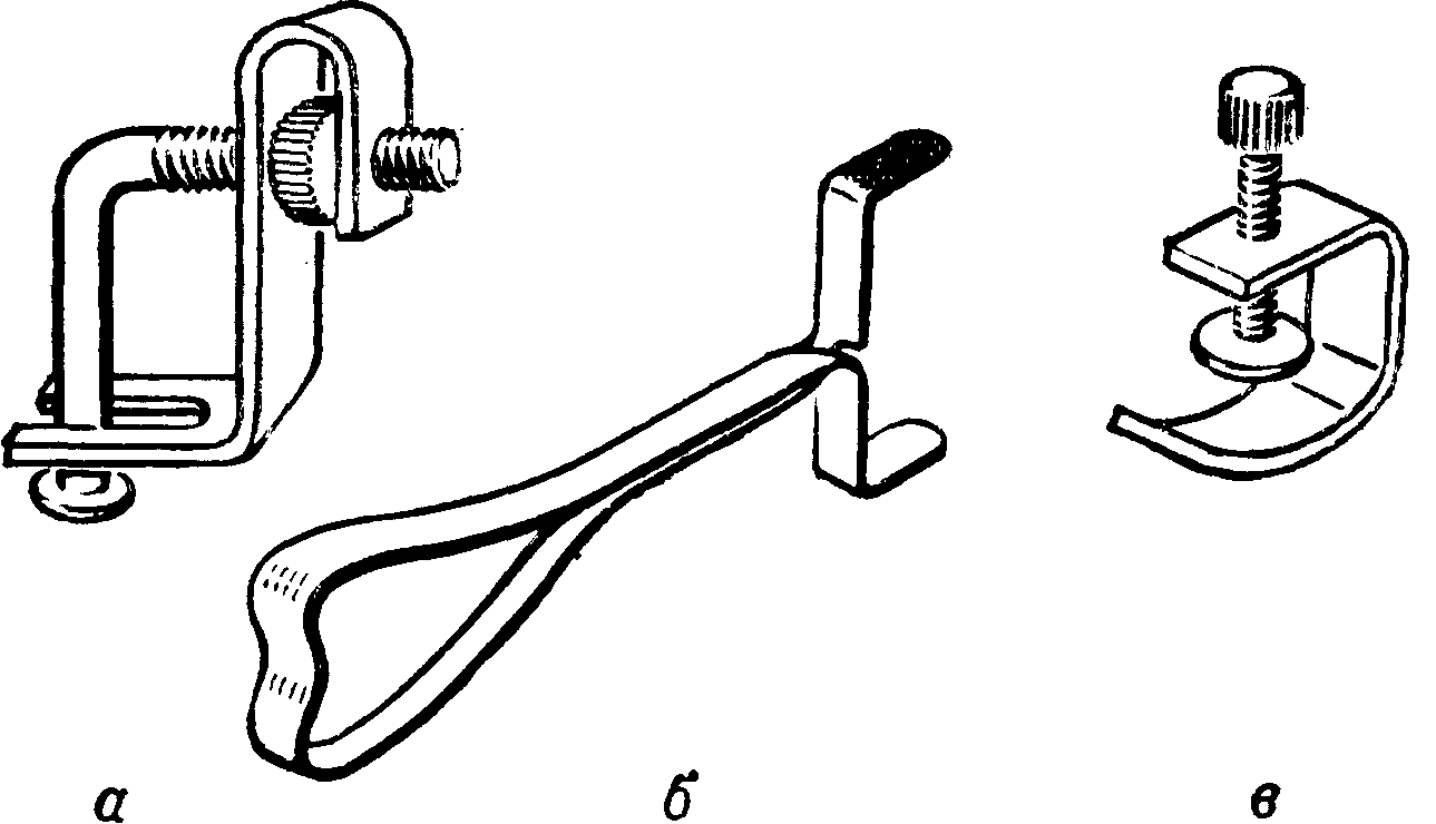
Clips - Holders... To attach the surgical linen to the skin during surgical operations, clamps are used, which are a hinged clamp with a ratchet; working jaws of the clamps are sharpened and easily pierce linen and skin (fig. 2, 29). To attach the surgical linen to the peritoneum, use hinged clamps with a denticle according to Mikulich (Fig. 2, 30) or plate clamps-clamps. For grasping and holding tissues, removing foreign bodies, introducing tampons into wounds, forceps are widely used - straight or curved (Fig. 2, 31). To grasp the mucous membrane of the stomach and intestines use a clamp for the intestinal wall (Fig. 2, 32) Ellis, the working jaws of which have several sharp teeth. For grasping and holding organs, various kinds of fenestrated clamps are used: during operations on the gallbladder - a clamp for the gallbladder (Fig. 2, 33); to hold the pancreas during its dissection and subsequent stitching with the intestine during pancreatoduodenal resections - a special clamp for the pancreas (Fig. 2, 34). End clamps are also used to extract stones from the gallbladder, to grasp and hold the tumor in the nasopharynx, to hold the lung during surgery, etc. There are also various auxiliary clamps for elastic tubes coming from blood transfusion devices, suction machines etc. Figure 3 shows screw and spring clamps for elastic tubes; one of them (Fig. 3, c) is used mainly for blood transfusion.
, Urological instruments, Surgical instruments.Bibliography: Kabatov Yu. F. Medical tools and equipment, M., 1977; Krendal P.E. and Kabatov Yu. F. Medical commodity science, M., 1974.
Yu. F. Kabatov.
5.1. GENERAL AND SPECIAL SURGICAL
TOOLS
There are currently enough a large number of classifications of medical instruments and options for dividing it into groups. Surgical and dental instruments can be distinguished according to their purpose.
Surgical instruments are divided into two groups.
General surgical instruments- these are the instruments most often used in the clinic and used for basic manipulations. Quite often, these tools are multifunctional.
Special tools are instruments that are used only in certain areas of surgery. Quite often, the tools of this group are used only when performing a specific stage of any one operation.
General surgical instruments, in turn, can be divided into 4 subgroups depending on their specific purpose:
tissue separation tools: scalpels, knives, scissors, osteotomes, chisels, wire cutters, etc .;
instruments for stopping bleeding: ligature needles of Cooper and Deschamp, hemostatic clamps, clips and clamps for their application;
tools for connecting fabrics: needle holders, surgical needles, tweezers for the application of Michel's staples, staplers, instruments for bone sutures, etc.;
auxiliary tools:
To create an exposition: retractors, hooks, mirrors, etc .;
For holding and displacing organs: tweezers, lifters, probes, etc.
By the number of component parts, instruments can be divided into one-piece (usually forged or stamped) - scalpels, chisels, chisels, hooks, as well as prefabricated, which, in turn, can be non-articulated (tweezers, trocars) and articulated (clamps, needle holders, forceps). The latter group is classified by the number of pivots: single-pivot (clamps, scissors, most forceps) and multi-pivot (double-gear nippers, gastric pulps).
In addition, according to technical specifications(GOST 19126-79), surgical instruments are divided into:
Sharp sharpened tools (sharp-cutting, cutting, stabbing);
Tools with spring properties (ratchet, hingeless);
Plate tools (hooks);
Wire tools (probes, some types of hooks, conductors);
Tubular instruments.
This division can be carried over to special instruments.
Dental instruments, among other things, are divided by purpose into:
General-purpose tools (dental burs, dental mirror, tweezers, scissors, probes);
Therapeutic instrumentation (for filling, treatment of periodontal diseases, etc. - floats, curettes, needle files, hooks);
Surgical instrumentation (for tooth extraction, treatment of periostitis, etc. - forceps, curettage spoons, elevators);
Instrumentation for endodontics.
All dental and general surgical instruments are combined into special sets according to the final purpose, for example, a set for the extraction of teeth (a separate set for the extraction of teeth in children, similar to smaller ones), a set for examination, a set for endodontics, a set for filling, etc. The composition of the kits varies somewhat depending on the medical institution, supplier, manufacturer, etc. Sometimes the habit of the dentist himself also influences the formation of the set. In this regard, it is advisable to highlight basic part of the set and additional.
It is quite natural that the basic set of tools should be of particular interest to the student.
The obligatory set of general surgical and special instruments is reflected in the “Appendix? 9 "to the order of the Ministry of Health and Social Development of the Russian Federation dated 04.14.06? 289 "On measures to further improve dental care for children in the Russian Federation."
5.2. TYPES OF SURGICAL INSTRUMENTS
5.2.1. Tissue Separation Tools
Scalpel- a small one-piece tool with a short blade and a long handle (Fig. 5.1). Designed for dissection of soft tissues (skin, subcutaneous tissue, aponeuroses, muscles, etc.). There are several types of scalpels: pointed, abdominal, eye. The latter differs only in smaller dimensions and, as a rule, is made of the pointed type. The use of a scalpel depends on the shape of its blade: the abdominal scalpel is used to cut the skin, the pointed one is used for more delicate manipulations, when, in addition to the incision, it is necessary to make a puncture. An eye scalpel is used for small incisions. There are modifications of the scalpel with replaceable blades. Currently, disposable scalpels are becoming more widespread. For particularly thin facial incisions
12 3
Rice. 5.1.Scalpels: 1 - abdominal scalpel; 2 - pointed scalpel; 3 - eye scalpel; 4 - disposable scalpel; 5 - blade holder
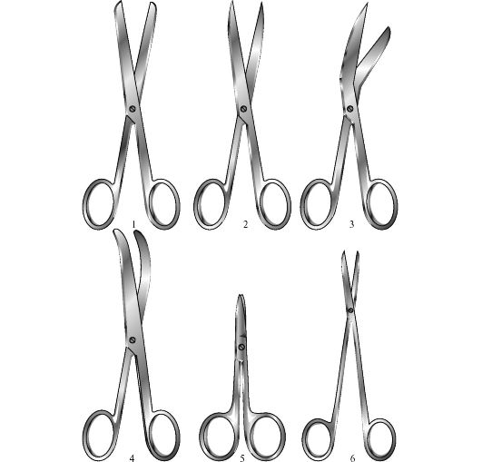
Rice. 5.2. Scissors:
1 - blunt scissors; 2 - pointed scissors; 3 - Richter's scissors; 4 - scissors curved along the plane (Cooper); 5 - eye scissors; 6 - vascular scissors
(for example, for plastic surgery) also use a microsurgical instrument - a blade holder. The cutting part of this tool is represented by a fragment of a razor blade, which allows, firstly, to make thinner and more accurate cuts, and secondly, to quickly replace the blade if necessary.
Scissorsare a collection tool. They consist of a working part (blades) and handles connected by a screw or rivet (Fig.5.2). The edges of the blades, closing, provide cutting of tissues. Scissors can be straight, curved in a plane or at an angle. In addition, there are blunt-pointed and pointed scissors. Small scissors, both straight and curved, are called eye scissors. The use of scissors depends on their shape, since scissors, in addition to cutting, also produce unwanted crushing of tissues. They are used where, for some reason, it is impossible to use a scalpel (for example, when cutting loose tissues or when it is necessary to make an incision to a certain depth without affecting the underlying layers). Typical is the use of straight and curved scissors to form a patch from a filmy plastic material (fascia, greater omentum, synthetic film).
Angled scissors (Richter's scissors) are typically used to dissect the peritoneum and pleura during laparotomy and thoracotomy, as well as to dissect the hernial sac. There are also modifications of scissors for dissecting gauze (with a thickened cutting part), plaster (with a beak at one end) bandages, as well as for longitudinal dissection of blood vessels. Microsurgical scissors are gradually gaining widespread use.
Chiselsand chisels are one-piece forged or stamped one-piece tools (Figure 5.3). They consist of a pointed working part (straight or curved) and a handle. The chisel is characterized by a straight handle of a simple shape with a flattened end ("heel", or striking part). The chisel has a massive handle, hollow inside, without flattening. These instruments are intended for processing bone: with a chisel, you can scrape off excess bone tissue (with osteosynthesis or plastics), and using a chisel and a hammer, the surgeon cuts or cuts the bone. The largest, strongest chisels are also called osteotomes.
Bone spoons- one-piece instruments with a working part made in the form of a small spoon with pointed edges (see Figure 5.3). Used to remove bone residues during treatment
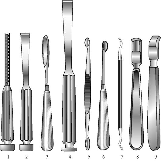
Rice. 5.3.Bone Surgery Instruments
Chisels and chisels: 1 - grooved chisel; 2 - straight bit; 3 - chisel Voyachek grooved; 4 - osteotome. Bone spoons: 5 - Volkmann's spoon; 6 - Bruns spoon; 7 - dental excavator. Raspators: 8 - straight Farabef raspatory; 9 - curved Farabef raspatory
multiple fractures or osteomyelitis. In addition to bone trays in dentistry, dental excavators are used, which are designed to remove temporary fillings, remove sequesters, cleanse the tooth cavity, etc.
Raspatoryare designed to remove the periosteum from the bones (see Figure 5.3). They consist of a working part - a cutting edge with a support platform and a strong handle. They can be straight and curved in shape.
Bone forcepsused for biting bone fragments - when treating wounds of the brain and facial parts of the head (Fig. 5.4). Luer pliers are distinguished by the rounded shape of the working part with a cavity inside, into which the bitten off bone fragment is placed. The Liston pliers are made as side cutters and provide a relatively thin and straight cutting line. To increase the cutting torque, a double gear is installed in the nippers. Dahlgren's nippers are distinguished by the fact that the cutting part is made in the form of a hook and can be replaced in case of breakage or wear. These forceps are used for trepanning of the bones of the cranial vault.
Rice. 5.4. Bone forceps:
1 - Liston nippers; 2 - curved Luer pliers; 3 - the appearance of the double transmission of the pliers; 4 - Dahlgren nippers
Tooth extraction forceps: each tooth corresponds to quite definite forceps, differing in numbers (Fig.5.5).
Elevatorsdental - designed to remove the remnants of tooth roots.
5.2.2. Instruments for stopping bleeding
To stop bleeding, various types of hemostatic clamps are mainly used (Fig. 5.6).
Hemostatic forceps consists of a handle with a rack and a working part. In this case, the shape and size of the working part can be different. Straight clamps are distinguished according to the shape of the working part.
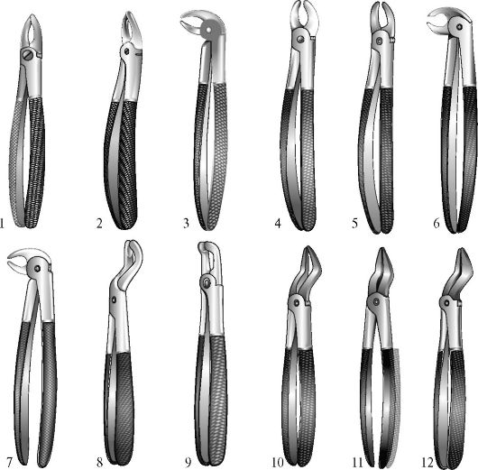
Rice. 5.5.Teeth extraction forceps (from: V.I. Bezak, 1969): 1 - straight? 2 - to remove incisors, canines and premolars; 2 - S-shaped М7 - to remove premolars; 3 - beak-shaped with converging cheeks? 13 - to remove roots; 4 - S-shaped? 17 - to remove right molars; 5- S-shaped? 18 - to remove left molars; 6 - beak-shaped (coronal)? 22 - to remove molars from both sides; 7 - beak-shaped with rounded non-converging cheeks? 33 - to remove teeth and roots; 8 - bayonet? 67 - to remove wisdom teeth; 9 - beak-shaped horizontal? 79 - for the removal of wisdom teeth with difficulty opening the mouth; 10 - bayonet (bayonet) with narrow cheeks? 51a - for removing roots and teeth with a destroyed crown; 11 - bayonet-shaped (bayonet) with middle cheeks? 51 - for removing roots and teeth with a destroyed crown; 12 - bayonet (bayonet) with wide cheeks? 52 - for removing roots and teeth with a destroyed crown
Rice. 5.6.Instruments for stopping bleeding. Hemostatic clamps: 1 - straight Billroth clamp; 2 - straight Kocher clamp; 3 - "mosquito" type clip; 4 - Hepfner's vascular clamp. Ligature needles: 5 - Deschamp ligature needle; 6 - Cooper's ligature needle
and curved. Curved clamps are more convenient; The Hepfner vascular forceps can be used to suture a damaged carotid artery end-to-end.
Ligature needlesare used to ligate the vessel throughout (see Figure 5.6). In maxillofacial surgery, they are used to ligate the carotid arteries and their branches. The tip of the needle may be sharp or blunt. In this case, the Cooper's needle is used to ligate a deeply located vessel, and the Deschamp's needle is used for a superficial one.
5.2.3. Tissue Joining Tools
Needle holdersin shape they closely resemble hemostatic clamps, but differ in a thicker and shorter working part. Designed to hold surgical needles
Rice. 5.7. Needle holders:
1 - Gegar needle holder; 2 - Troyanov's needle holder; 3 - Mathieu needle holder; 4 - microsurgical needle holder
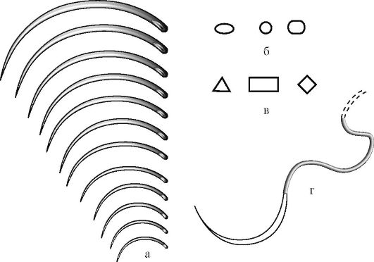
Rice. 5.8.Surgical needles:
a - surgical needles of different sizes. b - section of piercing needles; c - section of cutting needles; d - atraumatic needle with thread
in the process of suturing soft tissues. Microsurgical needle holders are used to work with thin sutures and small needles (Figure 5.7).
Surgical needles are intended for suturing fabrics (fig. 5.8). According to the shape of the needles are divided into straight and curved. On the cross-section, piercing (circular) and cutting (triangular, rectangular, in the shape of a trapezoid) are distinguished. By size, the needles are divided into 12 groups in length (numbers from 1 to 12, with the larger the number, the smaller the needle) and into 3 groups in thickness (thick, thin, eye). In addition, a separate group is made up of atraumatic needles, in the back of which sterile suture material is attached.
5.2.4. Supporting tools
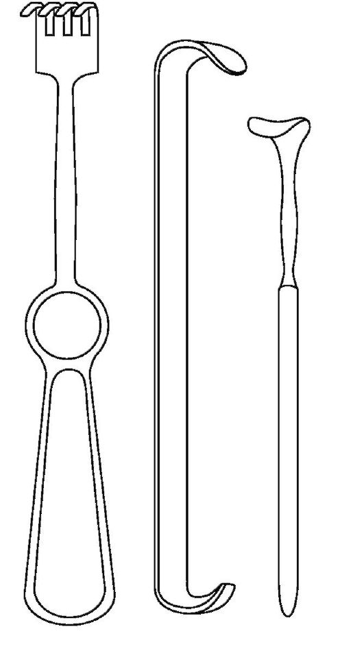
Hookscan be one-sided and two-sided (fig. 5.9). One-sided hooks consist of a handle and a working part. An example of these are Volkmann's three-pronged and four-pronged surgical hooks. The double-sided hook (Farabefa) is more versatile, as it has two working parts of different sizes. In addition, it injures the retained tissues less. The Farabef hook can be made in two versions - C-shaped and S-shaped. The saddle hook is used to hold the isthmus of the thyroid gland during operations on it and the trachea. The Limberg hook is used in the treatment of fractures of the zygomatic arch.
Gag (Fig.5.10) is intended for forced opening of the mouth in emergency conditions, some
12 3
Rice. 5.9. Hooks:
1 - four-pronged Volkman's hook;
2 - lamellar hook Farabef;
3 - small saddle hook
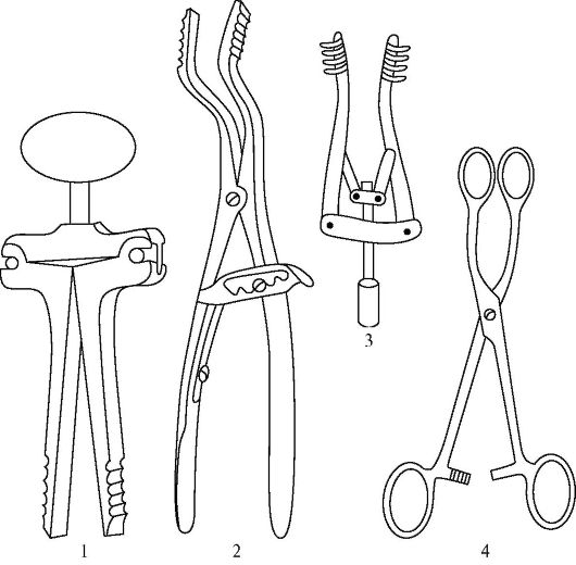
Rice. 5.10.Mouth dilators and retractors:
1 - screw rotor expander; 2 - rack-and-pinion retractor; 3 - screw retractor; 4 - language holder
modifications are used in a number of clinics for any interventions in the oral cavity.
Retractors (see Fig. 5.10) are used to expand the edges of the surgical wound. In maxillofacial surgery, small retractors with a screw thrust are widely used, for example, Edson's retractor.
Language holder(see Figure 5.10) is used to grasp the tongue and move it out during operations on the oral cavity.
Tweezers- two-piece tools with springy working parts (Fig. 5.11). Designed to grip and hold tissues, organs, dressings. Anatomical tweezers are distinguished by a more gentle, but at the same time less strong grip - they are held
Rice. 5.11.Tweezers and forceps:
1 - anatomical tweezers; 2 - surgical forceps; 3 - dental tweezers; 4 - eye forceps; 5 - forceps
easily injured anatomical formations. Surgical tweezers have teeth on the working surface that injure the tissues, but they grip them very tightly. Dental tweezers are curved, they are comfortable to work during the treatment of dental diseases - they are injected with turunda into the carious cavity. Eye tweezers are smaller and can be used for small manipulations.
Kornzangivary in size (large, medium, small) (see Figure 5.11). They can be straight and curved in shape. In appearance, they resemble hemostatic clamps, but have a stronger working part with a thickening at the end. They are designed to grab sterile linen, suture material, instruments from sterilizers. Quite often, curved forceps are used in the primary surgical treatment of wounds for their mechanical cleaning.
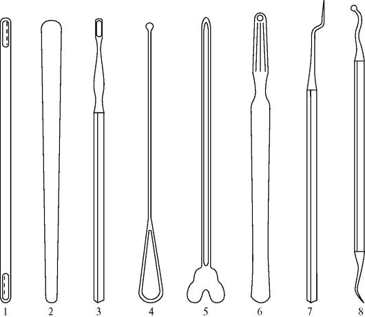
Rice. 5.12. Spatulas and probes:
1 - neurosurgical spatula; 2 - therapeutic spatula; 3 - dental spatula; 4 - bulbous probe; 5 - grooved probe; 6 - Kocher's probe goiter; 7 - dental bayonet probe; 8 - dental excavator
Spatulas- tools with a flattened and blunt working part (Fig. 5.12). Neurosurgical spatulas are used to push back brain tissue during neurosurgical interventions. A therapeutic spatula is needed to move towards the tongue when examining the oral cavity and the condition of the tonsils. Dental spatulas are mainly used for kneading filling paste.
Probes(see figure 5.12). The main purpose of the bulbous probe is the revision of the fistulous passages. A grooved probe is used when dissecting the fascia or aponeurosis to protect the underlying tissue from damage. The Kocher probe is used in a similar way to ligature needles for thyroid surgery. Dental
probes are used for revision of teeth - revealing softening of dentin, depth of carious cavity, etc. Dental excavators can be used for removing food debris, replacing fillings, scraping out granulations.
5.3. SPECIALIZED KITS
INSTRUMENTS
General surgical kit used in the primary surgical treatment of head and face wounds, treatment of purulent diseases, etc. Also, this set is an integral part of most specialized kits for plastic surgery, vascular surgery, oncosurgery, etc. The set consists of scalpels, hemostatic clamps, tweezers, probes, hooks (or retractors), scissors, needles and needle holders, forceps.
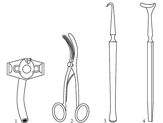
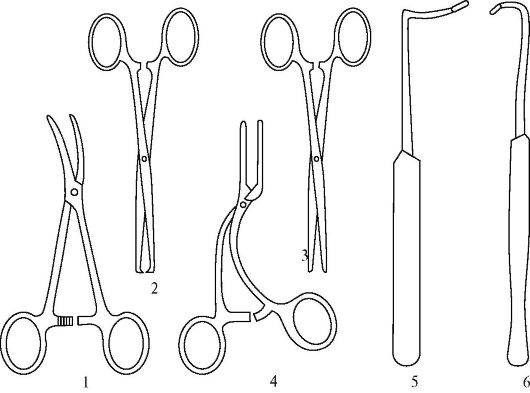
Rice. 5.13.Tracheostomy kit:
1 - tricyceostomy Luer cannula; 2 - Trusso's tracheostomy retractor; 3 - a sharp one-toothed hook for holding the trachea; 4 - saddle hook
![]()
Rice. 5.14.Basic set for dental examination: 1 - dental mirror; 2 - dental probe (angular); 3 - dental tweezers; 4 - dental spatula; 5 - dental excavator
Tracheostomy kit (Fig. 5.13) consists of a Trousseau tracheostomy retractor, a tracheostomy cannula, a sharp hook for holding the trachea and a saddle hook for the isthmus of the thyroid gland. The use of this kit is impossible without general surgical instruments.
The basic dental examination toolkit (Figure 5.14) includes:
- dental mirror- consists of a handle and a mirror itself - a curved mirror plate with a focal length of 75 mm. There are non-collapsible and collapsible modifications, and the collapsible mirrors are more convenient - the handle for some of them is made of plastic and fits better in the palm of your hand. Intended
mirrors for examining hard-to-reach areas of the oral cavity and the back-inner surfaces of the teeth;
- dental probe- there are different options executions - curved, sickle, bayonet. The probe is designed for the detection and revision of carious cavities;
- dental tweezers- intended for the introduction of turunda and tampons into a wound or carious cavity;
- dental spatula- can be two-sided or one-sided. It is used to knead the filling compound and grind it. Collapsible spatulas with replaceable tips are very convenient. Some manufacturers complete these kits with disposable tips and reusable pens that fit more comfortably in the palm of your hand;
- dental excavators- differ in the diameter of the spoon (from 0.7 to 2.4 mm, numbers from 1 to 4, respectively), are designed to extract fragments of hard dental tissues, food debris from the carious cavity, remove temporary fillings and dental deposits, scraping out granulations;
- trowels, corkscrew-trowels- differ in the size of the working parts, are intended for smoothing the filling mass when closing the carious cavity.
In addition, the basic set includes a kidney-shaped tray, in which instruments and dressings are placed. However, a number of manufacturers complete the basic kits with their own modified trays or cassettes for holding the instrument. Such cassettes greatly facilitate the work of a dentist.
A set of tools for filling (Fig.5.15):
- drill sleeves- are used to transfer torque from the stationary engine of the drill to the working part. If not a mechanical, but a pneumatic propeller is used, flexible twisted hoses and air ducts are used instead of hoses;
- handpieces for hose drills- straight and angular are available, they are used to fix rotating tools (burs, cutters, etc.);
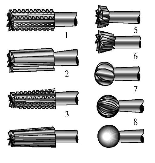
- dental burs serve for mechanical processing hard tissues (tooth, hardened filling mass), differ in shape and purpose: burs - for primary processing, finishers - for finishing, polishers - for polishing (Fig. 5.16);
- matrix for contour fillings used to temporarily strengthen the filling in case one of the walls of the tooth is missing.
A set of instruments for working with a root canal (Fig.5.17):
- root drills(manual and machine) - designed for expansion and alignment of root canals, differ in diameter (from 0.25 to 0.45 mm, numbers from 1 to 5, respectively);
- drilbor manual and machine - used for probing, expansion and filling of the root canal;
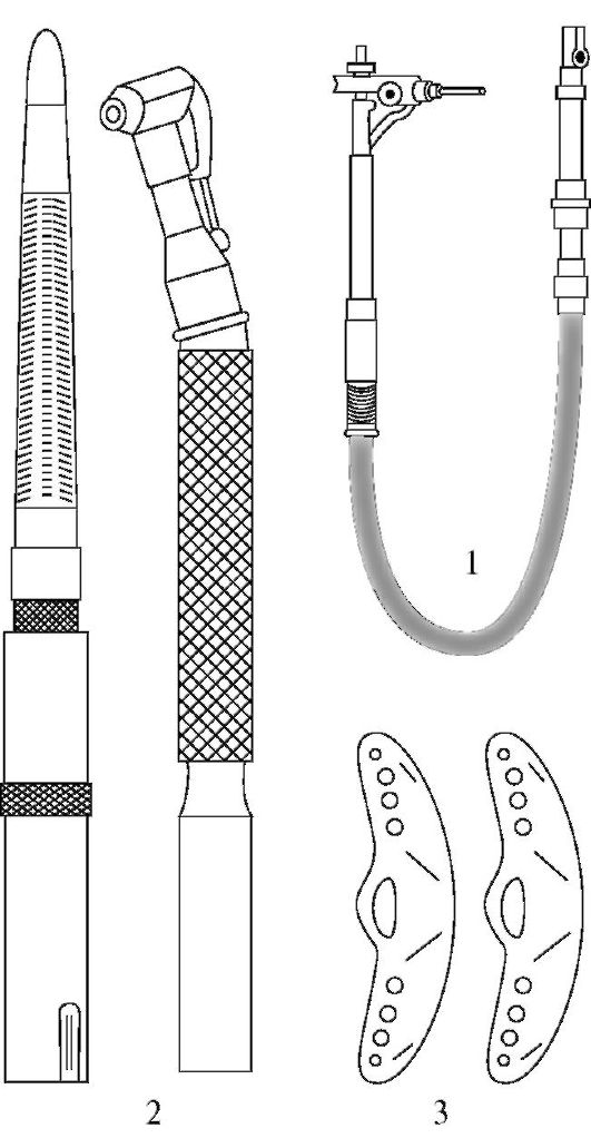
Rice. 5.15.Filling tools:
1 - drill sleeve; 2 - handpieces for a straight and angle drill; 3 - matrix for contour fillings
4 9
Rice. 5.16.Dental burs (from: V.I. Bezak, 1969). For the treatment of deep carious cavities: 1 - fissure cylindrical with double cutting; 2 - fissure cylindrical with single thread; 3 - fissure conical with double thread; 4 - fissure conical with double grooves. For shikolevoid processing; 6 - wheel-like back conical. For the treatment of shallow carious cavities: 7 - spherical (spherical) boron. To prepare the filling for polishing: 8 - spherical finish. To polish the filling: 9 - ball polisher
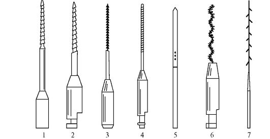
Rice. 5.17.Tools for working with a root canal: 1 - manual root drill; 2 - machine root drill; 3 - manual drill; 4 - machine drill; 5 - root needle; 6 - filler; 7 - pulpoextractor
- root needlesMiller - differ in diameter (0.17, 0.19, 0.21 mm, numbers 1, 2, 3, respectively), are intended for the introduction of medicinal substances into the root canal and its subsequent filling;
- canal fillers - designed to fill the root canal with a filling mass;
- pulp extractors- can be made with a long or short handle. Used to remove residual pulp from the root canal.
5.4. INSTRUMENT POSITION ON THE OPERATING SISTER'S TOOL TABLE
When performing general surgical operations, one should adhere to certain rules for the location of instrumentation and soft equipment.
The operating nurse covers the movable small instrument table with a double-folded sheet so that one half of the sheet covers the table, and the other hangs down and then
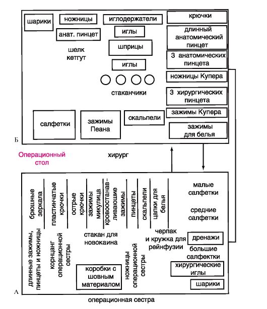
Rice. 5.18.Methods for placing instruments on a small instrument table: A - left-sided; B - right-sided
could close the collected tools. The operating nurse shifts the forceps necessary tools from a large instrument table to a small one and places them in a certain order. Depending on the position of the table - to the right of the operating table (next to the surgeon) or to the left of the table (next to the first assistant) - the instruments are placed in two ways.
The layout of the surgical team and the instrumental table for various operations is shown in Fig. 5.18.
With the position of the nurse's table to the right of the patient, the layout of the instruments and material is shown in Fig. 5.18, B. On the right edge of the table (from the side of the operating nurse) there are napkins in three packs: large napkins at the back, then medium and small. On the front edge (closer to the surgeon) there are instruments that are constantly needed during the operation: hemostatic clamps, Mikulich clamps, tweezers, scissors, hooks. The surgeon and his assistants take the instruments from the front edge of the table, and the nurse only maintains order on the table and restores the correct placement of the instruments.
The rear edge of the table is at the full disposal of the operating room nurse, and the surgeon does not touch it. Here are the spare tools ready for use; here is the suture material and scissors of the operating nurse.
The operating nurse is not allowed to touch instruments that have been in the operation and are stained with blood. As a last resort, she removes them with a forceps. With the position of the sister's table to the left of the patient, the layout of the instruments and material is shown in Fig. 5.18, A.
5.5. SURGICAL STAPLERS
For the first time the idea of using metal brackets when performing a gastric resection was expressed in 1903 by the Hungarian surgeon Hultl. The first apparatus for such manipulations was proposed in 1921 by the surgeon Petz. However, due to significant shortcomings, it was not widely used. A real breakthrough was made in 1949, when an apparatus for applying a circular vascular suture was developed and introduced into clinical practice in the USSR. In the 50-70s of the twentieth century in our country were developed and in different years more than 40 types of various staplers for connecting or suturing various organs were produced
and fabrics (Figure 5.19). Apparatus for applying a circular vascular suture (ASTs-4, ASTs-8, ASTs-20), a universal apparatus for suturing blood vessels (US-18), an apparatus for a linear vascular suture (ALSh-20), an apparatus for suturing the ear of the heart (UUS-20), devices for suturing the stump of the bronchus (UKB-25, UKB-16) and the root of the lung (UKL-40 and UKL-60), lung tissue (UTL-105), for stitching the bronchi (SB-2 and SB-3), an apparatus for applying an esophageal-gastric anastomosis (PCS), an apparatus for applying a gastrointestinal anastomosis (NZhKA-60), apparatus
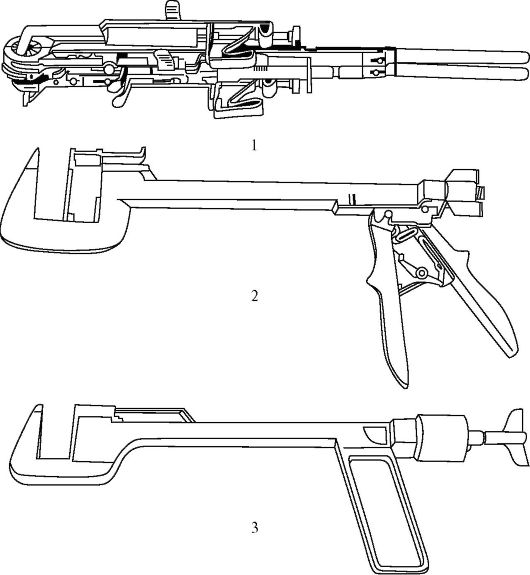
Rice. 5.19.Sewing machines: 1 - ASTs; 2 - UKL; 3 - UO
for suturing the sternum, ribs, collarbone and lower jaw (SGR-20, SRKCH-22). The apparatus for stitching soft tissues (SMT-2), devices for suturing organs (UO-40, UO-60) are multipurpose. In recent years, disposable staplers have become widespread.
The principle of the device of all these devices is the same. Each such apparatus consists of: a device for fixing the organs and tissues to be sutured; store with brackets; devices for ejecting staples; matrices for folding staples.
The essence of the stapler is as follows. After fixing the stitched tissues, the mechanism for pushing out the U-shaped staples is activated, which, having passed through the edges of the tissues, abut against the matrix and take a B-shape. Mechanical seam depending on the need, it can be one or two-row, linear or circular, with a longitudinal or transverse arrangement of brackets, superimposed simultaneously with all brackets or sequentially.
Suturing devices allow performing three typical surgical techniques: joining tissues or parts of organs, forming anastomoses of hollow organs, and forming organ stumps.
Staplers significantly reduce the dependence of the quality of seams on individual professional features surgeons, simplify the technique of the operation, speed up the operation, increase the reliability of the seams.
At the same time, there are contraindications to the use of stapling devices, primarily the presence of pathological changes in the stitched tissues (inflammatory or sclerotic processes).
5.6. DENTAL EQUIPMENT
Dental equipment is a set of devices necessary for the functioning of a dental office. The main devices are a dental chair, a drill, a dental illuminator, and a compressor. Equipment also includes diagnostic and display systems.
A dental chair is required to accommodate the patient. In some cases, it can be replaced by a regular chair (for example, during field preventive examinations).
The drill is necessary to create torque for working with burs, drillers, thin sections. In this case, the torque itself is transmitted using a flexible hose, or drive. A handpiece for the drill is located at the end of the drive. It can be straight or angular, depending on the damage to the tooth, its location, etc. The tip serves to secure the actual tool (see drills, drilburs).
A dental illuminator is required to illuminate the oral cavity. It consists of a support (stand), a lamp and a reflector. There are shadowless modifications of illuminators, which have several lamps located in a circle. The main requirement for illuminators is a clear limitation of the light field, since otherwise the light can hit the patient's eyes.
The compressor is used to dry the mouth, suction saliva, etc. It can be used to generate torque (when using special air hoses and air tips), and then it can act as a drill.
Diagnostic systems are designed, on the one hand, to determine the volume and type of treatment, and on the other, to objectively assess the results of treatment by both the doctor and the patient (imaging systems). These systems include X-ray diagnostic devices, negatoscopes, dental video systems.
X-ray machines are X-ray machines (classical or film), computed tomographs, NMR tomographs. Currently, the use of radio visographs is becoming more and more relevant, in which X-ray radiation is recorded not by film, but by a digital sensor connected to a computer, which performs the final processing and presentation of the image. There are panoramic and sighting radiovisographs. The use of computer technology in the work of a dentist can greatly facilitate the archiving of patient data by creating databases. As a result, previous images can be requested at any time for comparison with subsequent ones.
Negatoscopes are designed to facilitate the assessment of radiographs. In some cases, they are integrated into the treatment unit of the dental unit.
Dental video systems are an intraoral camera connected to a computer. The main requirements
required for such cameras are diminutiveness and hygiene.
Currently, dental equipment is fully or partially integrated into dental units.
A dental unit is a complex of electrical, mechanical and hydraulic elements that converts external energy into the energy of dental instruments and is designed to provide the necessary conditions for dental treatment. Note that, in accordance with the definition, the complex of the above
devices that are not even connected to each other can be considered a dental unit. However, hereinafter, industrially manufactured dental units will be described, made in the form of a single complex, all elements of which are interconnected (Fig. 5.20).
Classification of dental units
By mobility: portable autonomous, portable plug-in, mobile, stationary.
By completeness: complete, incomplete (one or more elements are missing).
By the number of instruments: for 1, 2, 3, 4 instruments and modular with the ability to connect instruments one by one.
By the type of illumination on the arms: without illumination, with one optical fiber, with illumination on several arms.
By the type of micromotor: air, electric without illumination, electric with illumination.
According to the system of evacuation of fluid from the oral cavity: with a saliva ejector, with a saliva ejector and an injection vacuum cleaner, with a saliva ejector and a vacuum cleaner.
Rice. 5.20.Exterior view of the dental unit (Smile unit)
The basic equipment of the dental unit includes:
Patient chair - can be hydraulically or electrically driven, in modern installations it allows, if necessary, to position the patient lying down or in the emergency position - the Trendelenburg position (with the head end lowered). The upholstery of the chair must be durable and easy to clean.
All surgical instruments are divided into general and special ones: general surgical instruments are used for surgical interventions in any anatomical areas. Special surgical instruments, as a rule, are instruments of the same purpose as general surgical instruments, but are designed to perform operations in "narrow" areas of surgery: thoracic, cardiovascular surgery, neurosurgery, gynecology, urology, ENT and maxillofacial surgery, as well as for endoscopy and endovideosurgery.
4.1 Classification of surgical instruments
All surgical instruments are conventionally divided into the following groups:
1) disconnecting;
2) exciting;
3) piercing;
4) expanding and pushing back;
5) probing;
6) auxiliary;
7) mechanized.
The vast majority of instruments are named after their creators.
4.2 Characteristics of their individual types
I. Tissue Release Tools
The main tools for severing tissue are cutting. Instruments for severing tissue include scalpels, amputation and resection knives, scissors, saws, etc.
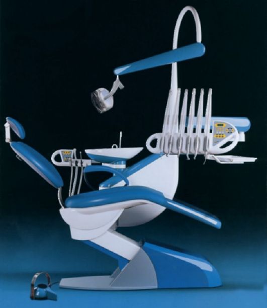
Scalpel - a sharp-edged surgical instrument (Fig. 1) used to sever soft tissues.
There are general surgical scalpels and special ones (ophthalmic, neurosurgical, etc.). General surgical scalpels can be solid-stamped and with removable blades. The scalpel has a handle and a blade; on the blade, a point, a back and an abdomen are distinguished. General surgical all-stamped scalpels are available in two types: spiky and abdominal(Fig. 1).
Fig. 1 Scalpels: a) pointed, b) abdominal
Surgical knives- Sharp-sharpened instruments designed to separate soft tissues during amputations, operative access to the organs of the chest cavity, etc. An incision made with a sharp knife is less painful and heals better.
Amputation knife intended for dissection of soft tissues during limb amputation. (Fig. 2, a).
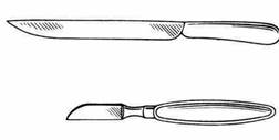
Fig. 2 Small amputation knife (a) and resection knife (b)
Resection knife(Fig. 2, b) is intended for dissection of dense tissues (small bones, more often phalanges) during hand and foot amputations, as well as during osteoplastic operations (joint resection, etc.).
Cartilaginous knife intended for separation of costal cartilage and sternum, as well as fibrous tissue.
Surgical scissors refer to cutting tools with a sharp sharpening, having two blades, dissecting tissue in the opposite motion (Fig. 3). Depending on the nature of this movement, distinguish articulated scissors(shearing action - along the blade) and guillotine shears(dissecting from top to bottom). Articulated scissors are used to separate soft tissues and dressings, dressings. Guillotine scissors are used to separate dense tissues (bones, cartilage, etc.).
Like other surgical instruments, scissors can be horizontally curved, i.e. in the plane of the table and vertically curved, which are more common.
Blunt scissors straight (Fig. 3, a) and curved (Fig. 3, c) (Cooper's) are most often used by surgeons to separate tissues, both on the surface and in the depth of the wound. Can be used to cut gauze. Curved scissors are used to sever adhesions in the pleural space or to separate organs from ligaments in the abdomen. They are also used to trim the ends of ligatures when closing a skin wound.
Pointed scissors straight and curved (Fig. 3, b) are used in cases when it is necessary to pierce the tissue before making the incision.
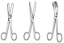
Rice. 3. Surgical scissors
Saws. In surgery, saws are used to intersect bones, which distinguish between three main types (Fig. 4): Charière's sheet saw (Fig. 4, c), Charière's arc saw (Fig. 4, b) and Gigli's saw (Fig. 4, a ), made in the form of a "sharp" wire, twisted into a spiral. In addition, traumatology uses different kinds electric saw.
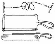
Fig. 4 Surgical saws: a) Charière sheet saw,
b) Charière's arc saw, c) Gigli's saw
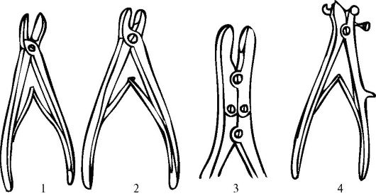
Surgical cutters used for biting bones (Fig. 5). Instruments in this category include Luer, Liston and Dahlgren's bony forceps, Doyen's rib scissors.
Disconnecting instruments (Fig. 6) include raspators (for separating the periosteum from the bone), chisels and osteotomes (for crossing bones - osteotomies), Volkmann's bone trays and others (for scraping the bone), trephins with a set of cutters (for drilling holes in bones).
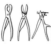
Fig. 5. Surgical nippers: a) Luer, b) Liston, c) Dahlgren, d) Doyen's rib scissors
Fig. 6. Disconnecting instruments: a) Farabef's raspator, b) chisels, b) bone spoons, c) trephine with a set of cutters
II. Grasping tissue tools
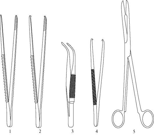
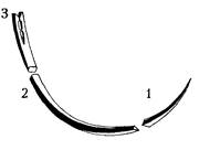
Hemostatic clamps - are used for clamping a bleeding vessel (temporary stop of bleeding), for ligation of a bleeding vessel (final stop of bleeding) (Fig. 7).
Kocher Serrated Hemostat - can be straight or curved, equipped with a lock (ratchet), and at the ends of the jaws there are teeth (two against one), and their entire working surface is covered with oblique notches.
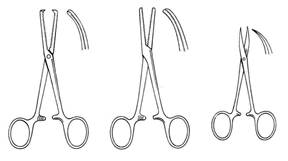
Fig. 7. Hemostatic clamps: a) Kocher, b) Billroth,
c) type "miskit"
Purpose:
1. Specially used to hold the reduced ends of the transected vessel in the thickness of the rough fibrous tissue (palmar and plantar aponeuroses, scalp, etc.).
2. To hold the superficial vessels of the thyroid gland (the initial purpose of the instrument).
3. For holding the dissected peritoneum and fibrous tissue (fascia and aponeurosis).
4. To hold the rib during the rib resection operation.
5. For holding and separating tissues during dissection during surgery.
Billroth's haemostatic forceps. It is similar in design to Kocher's hemostatic forceps. Differs in the presence of cross-cut jaws on the working surface. Can be with straight or curved cheeks (lips).
Purpose:
1. For ligation on a bleeding transverse vessel (less traumatic than Kocher's hemostatic forceps).
2. For holding the peritoneum or fixing it during dissection or suturing.
3. To hold the base of the appendix during appendectomy.
4. To perform blunt separation of tissues during surgery.
5. To open the abscess cavity and destroy the septa in the cavity.
Mosquito-type hemostatic forceps - short and light compared to the Billroth and Kocher hemostatic clamps, working jaws are distinguished by pointed ends, can be straight and curved;
Purpose:
1. For ligation of bleeding small vessels during neurosurgical operations.
2. For ligation with bleeding from parenchymal organs (liver, spleen, etc.), as well as in pediatric surgery.
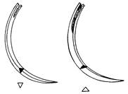
Vascular clips. They are intended for temporary application on the vascular legs of organs in order to stop blood circulation during an operation on the organ or during its removal (kidney, spleen, etc.) or for temporary application to the vessels when restoring their integrity (imposing a vascular suture) or restoring their patency (Fig. eight). Vascular clamps differ from hemostatic clamps in the figured structure of working sponges and a ratchet with a large number of teeth, which allows you to smoothly regulate the force of squeezing the vessel in order to injure the inner shell as little as possible. The configuration of the working jaws can be angular and arcuate (with different radius of curvature of the circle).
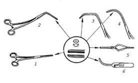
Fig. 8. Vascular clamps: 1-straight, 2-angled, 3-Satinsky clamp, 4- curved, 5-6 bulldog-type vascular clamps
Clamp for the renal pedicle of Fedorov- is a large and long clamp, curved along the plane. Used as a pedicle clamp near the hilum during nephrectomy (Fig. 9).
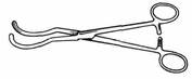
Fig. 9. Clamp for the renal pedicle of Fedorov
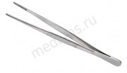
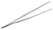
Tweezers - An instrument that is widely used in surgical practice and has a spring-loaded design, it is designed to grasp and hold a variety of tissues, materials and small instruments (Fig. 10).
The shape of the tweezers is straight or curved, depending on functional purpose... In special surgical instruments, special purpose tweezers are used.
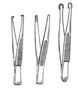
Fig. 10. Tweezers: a) surgical, b) anatomical,
c) serrated-clawed
Anatomical forceps(Fig. 10, a) has a transverse notch on the working surface of the jaws. It is used to hold easily injured organs and tissues of structures (peritoneum, vessel, nerve, intestine, etc.).
Surgical forceps(Fig. 10, b) is used to work with denser tissues (mainly skin, bone, etc.). Inevitably injures tissue.
Serrated tweezers(Fig. 10, c) has an extension in the form of a foot, on which there are notches (teeth). It has a greater fixation ability than surgical forceps, as it has a larger gripping area and a larger number of teeth. Designed to hold dense tissue (tendon, skin).
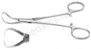
Clips for surgical drapes - hoes (Fig. 11, a) are intended for fixing sterile surgical linen (sheet, towel, etc.) to the patient's skin. In this case, only the operating field is opened for the surgeon, and the rest of the body must be covered with sterile linen (sheets, etc.). The slippers can replace other instruments when holding organs and individual anatomical structures (tongue, rib, spermatic cord, etc.).
The working jaws of these instruments are sharpened at the ends for a better grip of the surgical drape.
Clamp for attaching surgical drapes (Mikulich) to the peritoneum (Fig. 11, b) resembles Kocher's hemostatic forceps in design, but apart from the teeth it has an oblique cut on the working jaws.
Fig. 11. Clips for surgical drapes: a) underwear clip,
b) Mikulich clamp
Kornzang- a special clamp designed for the supply of sterile instruments and dressings, for the introduction of tampons and drains. Kornzang has oval sponges, on the working surface of which there is an oval depression and an oblique notch (Fig. 12).
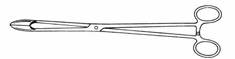
Fig. 12. Kornzang
Tissue holding clamps . In general surgery, tissue fixation clamps are used for a variety of purposes. Most often they are used to hold tissue firmly, but not to separate it from the surrounding tissue: for the purpose of producing traction (traction) or oppositions.
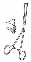
Fig. 13. Clamps for holding tissues: a) tissue clamp,
b) bullet forceps
To achieve the above goals, these tools are designed in such a way that the most important part of them are the ends of the working jaws, which are tightly pressed against each other, and there is a working space between the working jaws. Sometimes there are teeth that fix the instrument well, but they make it traumatic for tissues (Fig. 13, a).
Vaginal cervical forceps (bullet forceps)- the ends of the sponges are pointed (one tooth against the other), there is a ratchet (Fig. 13, b).
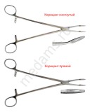
Gastric and intestinal pulp (clamps)
Atraumatic intestinal pulp- the ends of the working jaws have the form of transverse stripes, on the inner surfaces of which there are notches (Fig. 14, a). It is used to hold the intestinal wall during colostomy and gastrostomy operations., To stop bleeding when the source is not installed. It can also be used to hold soft and easily injured structures (fallopian tubes, ureter, appendix, etc.).
Fig. 14. Atraumatic intestinal (a) and hard gastric (b) pulp
Hard (crushing) gastric pulp of Payer's - superimposed on the removed part of the stomach during its resection (Fig. 14, b).
Needle holder - a surgical instrument designed to hold a surgical needle as it passes through tissue when suturing (joining tissue). The design of the needle holder is similar to a hemostatic clamp (Fig. 15).
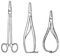
Fig. 15. Needle holders: a) Gegara, b) Troyanova, c) Mathieu
III. Tissue piercing instruments
Surgical needle is an indispensable tool for suturing and consists of three parts: an ear, a body and a tip (point) (Fig. 16).
Rice. 16. Components of a surgical needle: 1- tip (point),
2 - body, 3 - ear.
Straight needles, ski-shaped needles with a bend near the tip, and arcuate needles are distinguished in shape. Depending on the cross-sectional shape, surgical needles are round (oval), triangular, square, rectangular, trapezoidal (Fig. 17).
The purpose of the needles is different depending on the cross-sectional shape.
1. Round (piercing) needles are also called "intestinal" needles. They are used to pierce the walls of hollow organs: stomach, small and large intestine, biliary tract. These needles can also be used to suture vessels and nerves.
2. Triangular, or "cutting", needles connect the edges of dense organs and tissues - sternum, fascia, tendons, skin. One of the cutting edges of the needle body can be facing outward (curved-cutting needle) or inward (concave-cutting needle) (Fig. 18).
![]()
Rice. 17. Features of the shape of the cross-section of the needle body: 1 - round; 2 - oval; 3 - triangular; 4 - square; 5 - rectangular; 6 - trapezoidal.
A curved cutting needle is used to suture especially strong tissues (aponeurosis, tendon, scars, etc.). With this version of the cross-section of the needle body, the destruction of the inner edge of the channel created by the needle is excluded, and thread cutting is prevented. The concave cutting needle is used in many areas of surgery due to its versatility.
Rice. 18. Concave-cutting (1) and concave-cutting (2) needles.
3. Needles with square, rectangular and trapezoidal sections are used for suturing tissues in microsurgery, plastic and eye surgery.
Using needles different shapes depending on the level of actions in the wound, it obeys certain patterns.
1. Tissues located superficially, or organs brought to the surface of the body, can be sutured using straight needles. With such needles, for example, it is possible to suture the skin, the intestine removed from the abdominal cavity, the isolated tendon.
2. The closer the tissue is sutured to the bottom of the narrow wound, the greater the part of the circumference should be the needle.
3. When working in conditions of limited visibility and the need for constant monitoring in the field of view of the position of the needle tip at the most important anatomical elements (vessels and nerves), shortened surgical needles are used.
V modern designs atraumatic needles the thread and the body of the needle are a single whole (Fig. 19), which gives a number of advantages:
Rice. 19. Atraumatic needle
The diameter of the body of the atraumatic needle and the thickness of the thread are the same, minimizing damage to the stitched tissues;
An atraumatic needle is followed by an ordinary thread, as opposed to a double thread with an open or closed eye needle;
Suture material razvlechenie is excluded.
Infusion needle intended for subcutaneous administration of fluid. It has several lateral holes at the end. The needle for blood transfusion (Dufo), in addition to the olive-shaped part, has a corrugated square section on the head for ease of holding and injecting into a vein.
Needle "butterfly"(Strauss "a) short and thick, has a plate near the head, convenient for holding the needle during vein puncture and fixing during prolonged infusion.
Teardrop needle can be straight or curved. It is used to open a vein when a catheter is inserted.
Spinal puncture needle(Bier "a) is distinguished by a massive thick head, easy to hold, as well as a special design of the mandrel, which has its own head. The mandrin fits snugly into the needle channel and its cut coincides with the cut of the needle. Thus, the needle and mandrel make up a single pointed shaft, relatively easily piercing dense tissue surrounding the spinal canal.Most puncture-biopsy needles are arranged according to the same type.When the end of the needle reaches the required depth, the mandrel is removed and a syringe cone is inserted into the head of the needle, with which the required amount of contents is extracted.
Fig. 20. Endoscopic trocars
Trocar - a stabbing surgical instrument designed to puncture the walls of human body cavities in order to remove fluids, introduce endoscopic instruments, and also to take material (biopsy) (Fig. 20). The trocar consists of two parts: a rod (stylet) sharpened on one side with a handle on the other, and a tube (cannula). The cannula is shorter than the rod.
The rod, together with the cannula, is inserted through the skin and penetrates into the body cavity (peritoneal or pleural). The stylet is then removed and the tube remains in the cavity. Through it, a catheter is inserted for the outflow of contents (ascites, pleural empyema, etc.), as well as for the introduction of endoscopic devices and instruments.
IV. Expanding and displacing tissue tools
Instruments of this group are used for better exposure of the surgical wound after a skin incision, for pushing back organs and tissues in order to ensure prompt access and the best visibility of the operating field during the operation.
Retractors (hooks) - are used for superficial retraction: serrated (Volkmann, etc.) and lamellar (Farabef, etc.) or for deep retraction (mirrors), the working part of which is flat or saddle-shaped with a surface polished to a shine that reflects light, which is necessary for additional illumination of the operating room fields (Fig. 21).
Sharp hooks are used to hold the edges of skin wounds, aponeurosis, and other dense structures. Blunt hooks are applied to more delicate tissues (muscles, tendons, etc.).
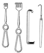
Fig. 21. Retractors (hooks): a) and b) Volkmann gears,
c) lamellar Farabef, d) sharp single-toothed
Volkmann serrated hook - has an all-metal handle or with a hole for a finger of various configurations; the working surface is represented by multi-toothed sharp or blunt hooks.
Farabef lamellar hook- is a plate with bent ends and a surface treated to a shine, serves to dilute the edges of the wound and soft tissues, to divert large blood vessels and nerves.
Mirrors. Wide and flat plate hooks are called mirrors. Abroad they are called retractors, just like hooks (Fig. 22). It is used for retraction of the abdominal cavity of organs (liver, spleen, etc.) during operations of cholecystectomy, vagotomy, lumbar sympathectomy, etc.
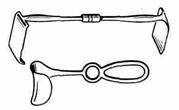
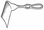
Fig. 22. Mirrors: a) angular and C-shaped, b) hepatic
Retractors - double-sided mirrors that do not require holding during the operation, because they are equipped with a self-locking device and a ratchet (Fig. 23).
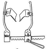
Fig. 23. Screw retractor
Shovels, elevators (lifts), spatulas for pushing back and moving apart various organs and tissues.
![]()
Fig. 23. Elevator (lift) (a), Buyalsky's spatula (b)
Dissectors - tools for moving tissue apart. These are the main tools for highlighting anatomical features in the area of the lung root.
V. Probing instruments
Probing instruments include probes(Fig. 24) , bougie, guides, catheters, cannulas... The most common probe is the grooved Nelaton probe (Fig. 24, a), which, like the Kocher probe, serves to dissect tissue along the groove or incisions. A bulbous probe is used to probe the cavities and ducts (Fig. 24, b).
![]()
Fig. 24. Probes: a) grooved Nelaton, b) Kocher
VI. Auxiliary tools
Ligature needle - it is an instrument with which a surgical thread (ligature) is passed under or through the anatomical structure on which the surgery is performed (Fig. 25). More often, a ligature needle is used to guide the ligature under blood vessels and ducts. Working part such a needle resembles a curved oval-section surgical needle, the eye of which is located at the beginning of a blunt (Deschamp's needle) (Fig. 25) or pointed end (Cooper's needle). In this case, the bending of the working part can be both to the right and to the left.
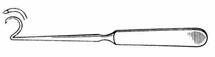
Fig. 25. Deschamp ligature needle
Vii. Mechanized tools
Mechanized instruments include automatic tissue staplers, cystourethroscopes, sigmoidoscope, bipolar forceps, fibroesophagogastroduodenoscope.
5. Technique of performing incisions with the SCALPEL.
Performing incisions of various shapes and sizes requires the surgeon different ways holding the scalpel (Fig. 26). The most comfortable position of the scalpel in the hand is provided when holding the instrument with three fingers (like a writing pen). This position allows precise and subtle movements. If it is necessary to carry out figured cuts or fine manipulations with a high degree of accuracy, the scalpel is held like a writing pen using a V finger support. In this case, the brush should rest on the two phalanges of the V finger or on the entire finger (as when writing with a pen), which allows you to more confidently and accurately manipulate the scalpel.
Holding the scalpel like a table knife is used when performing sufficiently deep straight long cuts when a certain pressure on the scalpel is required (for example, various types of midline laparotomy).
The position of the scalpel like a violin bow is used in the production of linear incisions, where there is no need to press on the instrument (dissection of subcutaneous adipose tissue, dissection of fascia, etc.).
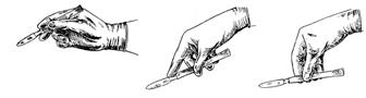
Fig. 26. The position of the scalpel in the surgeon's hand a) like a writing pen, b) like a table knife, c) like a bow
One of the main principles of making a skin incision is the same depth throughout its entire length. To achieve this goal, the scalpel at the starting point of the incision is set perpendicular to the plane of the skin and is injected like a spear to the depth of the planned incision. Then, tilting the instrument by about 45-60 °, the incision is continued in one even and smooth movement until the end point, where the scalpel is again brought to a vertical position relative to the skin. This technique also makes it possible to achieve the same length of the wound at the level of all dissected layers and to bring the size of the skin incision as close as possible to the volume of the surgical wound. When dissecting the skin, the scalpel should always be pulled towards oneself, starting from the outermost point of the incision. Sometimes the scalpel is pulled away from itself (for example, when dissecting the fascia with a fluted probe). When making an incision in the skin, it is necessary to ensure that it is perpendicular to its plane. The incision line should always be clearly visible to the surgeon. For complex incisions, it is advisable to first outline the skin incision line with a dye.
6. Physical methods separation of tissues
6.1 Plasma flow method (plasma scalpel)
For the separation of tissues in this case, a plasma flow is used, which is formed by passing a high-strength electric current through a high-speed jet of inert gas. The working part of the "plasma" scalpel is a metal cylinder with a pointed part and a nozzle.
The advantages of the plasma flow method are: high cutting speed of tissues due to the significant power of the flow, pronounced analgesic effect of the plasma flow, sterilization of the wound due to ultraviolet radiation and the release of atomic oxygen (ozone), achieving a hemostatic effect with a blood vessel diameter of no more than 1.5 mm (larger diameter vessels must be sutured or ligated), no damaging effect on the surgeon's eyes, the possibility of achieving a "biological welding" effect.
6.2 Cryosurgical method
The method is based on the possibility of removing a pathological formation after its rapid local freezing with a cryagent either in a spray mode or in a contact mode.
The working part of the cryosurgery devices are quickly cooled handpieces.
Cryagents are liquid nitrogen, freon, carbon dioxide in the form of dry ice, etc.
Local tissue freezing is one of the main methods of destruction in stereotaxic neurosurgery.
The cryosurgical method has found application in oncology, proctology (when removing a malignant tumor of the rectum), urology, etc.
6.3 Electrosurgical method (electric knife)
Disconnection of tissues in this way occurs due to the conversion of electrical energy into heat. For tissue dissection, an unmodulated electricity high frequency. Under the action of a high-frequency current, the continuous movement of ions in the tissues leads to the release of a significant amount of heat, which causes the evaporation of cellular elements (transition of liquid into gas) with the destruction of intercellular connections (separation of tissues). The emergence of "lightning" between the electrode and tissues is the main criterion for the correctness of performing electrosurgical cutting. Tissue dissection is more effective when the electrode has a sharp edge, providing maximum energy density.
6.4 Ultrasonic tissue separation
(ultrasonic cutting tools)
In ultrasonic surgery, instruments (knives, saws, drills) are used, the cutting edge of which continuously vibrates with a frequency of 10-100 kHz and an amplitude of 5-50 microns. To achieve these parameters, a magnetostrictive or piezoelectric phenomenon is usually used. High-frequency vibration provides tissue separation due to mechanical destruction of intercellular connections and the development of a cavitation effect (negative pressure formed in tissues due to the development of cavitation leads to boiling of intra- and intercellular fluid at a temperature of 38 ° C. The resulting vapor destroys the cell membranes and, spreading along intercellular spaces, separates tissues). The use of an ultrasonic knife is most advisable for the isolation and excision of scars, removal of tumors, opening of inflammatory foci, allows you to perform a kind of "soft" preparation - tissue stratification and separation of pathologically altered structures from normal ones.
Bone dissection (sternotomy, laminectomy, claviculotomy, etc.) is performed with an ultrasonic saw, on the cutting edge of which teeth are located with a step and a height of 1 mm.
6.5 Laser scalpel
The mechanism of action of a laser beam on biological tissues is based on the thermal effect on a limited area of the body with the energy of a monochromatic coherent light beam. In the "irradiated" place, the temperature can rise to 400 "C, providing instant combustion and evaporation of the pathologically altered area. The thermal effect on the surrounding tissues extends over a very short distance, since the diameter of the focused beam does not exceed 0.01 mm. Under the influence of laser radiation, occurs not only coagulation of proteins of living tissue, but also its "explosive" destruction during the instantaneous transition of tissue fluid into a gaseous state.
7. RULES FOR USE OF SURGICAL
INSTRUMENTS
All surgical instruments are tools for performing certain actions. To do this, they must be completely controllable, that is, they must be like an extension of the surgeon's hand. This is achieved only when correct position tool in hand. Of course, the “controllability” of most typical surgical instruments is ensured by the observance of the “rule of three fingers”. It is as follows. The instrument is held by I, II and III fingers of the hand: I and III are held with fingers, the branches are brought together and spread (if there are rings in the handle of the instrument, fingers are inserted into them); The second finger is a guide, it is placed over the instrument and guides the instrument to the desired object, in the required direction (Fig. 27). At the same time, as it was said, the surgeon must see his palm.
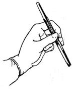
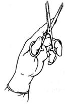
The tweezers also include the “rule of three fingers”, which is used to hold it like a writing pen (Fig. 28). The force of squeezing the tweezers with your fingers should be minimal, but not excessive, since soft tissues are easy to crush, dense ones to cut through, and hard ones are crumbled.
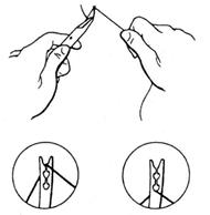
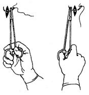
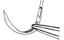
Fig. 30 Position of the needle in the needle holder
The needle is grasped with a needle holder, usually at the border of the middle and outer thirds of its length (Fig. 31). Depending on the direction of the puncture of the stitched tissues, the needle is fixed in the needle holder with the tip towards itself or away from itself, or to the left or to the right.
When suturing, measure the thickness of the thread and needle. The thread must be inserted into the second slot (Fig. 29) to ensure that it is firmly secured in the needle. The exception is thick threads, which are left in the first notch of the needle eye.
When sewing the fabric, rotational movements are made with the forearm in the direction of the needle point (Fig. 30). In this case, you should aim the needle in advance, determining the place of both its injection and injection. To ensure the puncture of the needle at the intended point, it is permissible to carefully insert the tissue onto the needle using open tweezers, tightly pressed by the ribs of the jaws on both sides of the intended place of puncture of the needle. This technique is useful when sewing very dense fabrics. If only the end of the needle appears when punching out, then you cannot grip it with the needle holder. It is necessary to intercept the needle closer to the ear and push it further. The withdrawn needle should not be taken with tweezers, but should strive to immediately grasp it with the needle holder. To do this, when applying a suture "on oneself", it is necessary to first pronate the forearm, which is in the supination position at the time of the needle sticking out. When applying a suture "from oneself" at the time of the puncture, the forearm is in the pronation position and, when removing the needle, it must be supined first. If you do not change the position of the forearm when removing the needle, then this will have to be done by rotating the shoulder. Sewing of tissues should be carried out with such pronation and supination movements of the forearm with a motionless hand fixing the needle holder.
8. SUTURE MATERIAL
Currently existing suture materials are classified according to several criteria.
By structure, the following types of threads are distinguished.
1. Monofilament (often incorrectly referred to by the outdated term "monofilament") is a single fiber with smooth surface... This type of thread includes such widely used materials as prolene, ethylon, dermalon, makson, nylon, surzhilen, surzhipro, miralen, dafilon, koralen (flexamide), maxilen, steel wire, etc. (Fig. 31, a)
2. A complex thread consists of many fibers (often surgeons call a complex thread multifilament, which is not recommended by modern standards). Depending on the method of connecting these fibers, three types of complex threads are distinguished (Fig. 31, b, c, d).
I. Twisted-fibers yarns are twisted along the axis, for example, flax, twisted silk.
2. Wicker - fibers are woven like a rope, for example, lavsan, etibond, mersilei, mersilk, nurolon, dexon II, etc.
3. Coated thread - braided thread impregnated and / or covered polymeric materials, for example, vicryl, polysorb, surzhidak, tikron, bralon, supramid, fluorex, fluorlin.
Fig. 31 Types of threads: monofilament (a), complex twisted (b), complex braided (c), complex with a polymer coating (d).
According to the ability to resorb (biodegradation) in the tissues of the body, three types of suture materials are distinguished:
Resorbable (absorbable) - catgut (simple, chrome-plated, with accelerated deadline resorption), materials based on polyglycolides (vicryl, polysorb, dexon, maxon), materials based on cellulose (occelon, cacelon, rimin), based on polylecaprone 25 (monocryl), polydioxanone, polyurethane, tendon threads.
Surgical Instruments.
Rice. 1. The scalpel is abdominal.
Rice. 2. The scalpel is pointed.
Rice. 3. Amputation knife.
Rice. 4. Resection knife.
Rice. 5. Scissors are straight pointed.
Rice. 6. Blunt scissors.
Rice. 7. Curved scissors.
Rice. 8. Scissors curved in a plane.
Rice. 9. Scissors with a narrow blade, curved in a plane.
Rice. 10. Scissors for cutting.
Rice. 11. Scissors with a button.
Rice. 12. Surgical forceps.
Rice. 13. Gripping tweezers.
Rice. 14. Tweezers anatomical.
Rice. 15. Straight hemostatic clamp with thread.
Rice. 16. Curved styptic clamp with groove.
Rice. 17. Hemostatic straight line clamp with thread and teeth.
Rice. 18. Curved styptic clamp with grooves and teeth.
Rice. 19. Hemostatic clamp of the "Mosquito" type with straight jaws.
Rice. 20. Hemostatic clamp of the "Mosquito" type with curved jaws.
Rice. 21. Hemostatic forceps for crushing tissues.
Rice. 22. Probe is thymus.
Rice. 23. The probe is bulbous with an eyelet.
Rice. 24. The probe is bulbous, two-sided.
Rice. 25. The probe is grooved.
Rice. 26. Lamellar hooks.
Rice. 27. The hook is blunt, two-toothed.
Rice. 28. The hook is blunt, four-pronged.
Rice. 29. The hook is sharp, two-toothed.
Rice. 30. The hook is sharp, four-pronged.
Rice. 31. Clamp for attaching surgical drapes.
Rice. 32. Clamp with ratchet for attaching surgical drapes.
Rice. 33. Clamp for attaching napkins to the peritoneum.
Rice. 34. Clamp with ratchet for attaching napkins to the peritoneum.
Rice. 35. Surgical skin needles.
Rice. 36. Surgical needles, steeply curved.
Rice. 37. Surgical intestinal needles, curved.
Rice. 38. Surgical intestinal needles, straight.
Rice. 39. Needle ligature blunt right.
Rice. 40. Needle ligature sharp right.
Rice. 41. Small needle holder with curved handle and ratchet.
Rice. 42. Needle holder with a curved handle and a large rack.
Rice. 43. Deep cavity needle holder.
Rice. 44. Michel metal brackets.
Rice. 45. Forceps for applying and removing staples.
Rice. 46. Autotweezers for applying metal staples.
Rice. 47. Brace with a set of cutters.
Rice. 48. Electrotrap.
Rice. 49. Nippers Egorov - Freidin.
Rice. 50. Powerful semicircular jaw pliers.
Rice. 51. Nippers are neurosurgical.
Rice. 52. Dahlgren Nippers.
Rice. 53. Universal retractor with a set of multi-toothed and flat jaws.
Rice. 54. Wire saw and handle to it.
Rice. 55. Guide for wire saws.
Rice. 56. Curved hemostatic neurosurgical forceps.
Rice. 57. Neurosurgical scissors, curved along the edge and along the plane.
Rice. 58. Curved neurosurgical forceps.
Surgical instrumentation... Rice. 59-97.
Rice. 59. Tweezers fenestrated for grasping brain tumors.
Rice. 60. Spatula neurosurgical.
Rice. 61. A sharp spoon for removing brain tumors.
Rice. 62. Bilateral neurosurgical curette for removal of brain tumors.
Rice. 63. Cannulas for puncture of the cerebral ventricles.
Rice. 64. Forceps for applying a clip to a magazine (frame).
Rice. 65. Neurosurgical aspiration tubes.
Rice. 66. A set of instruments for bipolar coagulation.
Rice. 67. Costal raspator.
Rice. 68. Costal scissors.
Rice. 69. Sternot.
Rice. 70. Rack-and-pinion wound spreader for the chest cavity.
Rice. 71. Wound retractor for the chest cavity, screw.
Fig .. 72. Mirror wire for the removal of the lungs and heart.
Rice. 73. Forceps for grasping the lung.
Rice. 74. Broncho-fixator.
Rice. 75. Clips straight and curved for the wedge-shaped lung.
Rice. 76. Mirror for taking the heart out.
Rice. 77. Clip for the ear of the heart.
Rice. 78. The commissure is the nail.
Rice. 79. Valvulot with two knives.
Rice. 80. Dilator for narrowed openings of the heart.
Rice. 81. Fork for lowering the ligature.
Rice. 82. Mirror for the abdominal wall.
Rice. 83. Mirror for liver abduction.
Rice. 84. Straight intestinal elastic forceps.
Rice. 85. Rigid intestinal clamp.
Rice. 86. Intestinal crushing pulp.
Rice. 87. Clamp for the gallbladder.
Rice. 88. Spoons for removing gallstones.
Rice. 89. Trocars.
Rice. 90. Rectal speculum.
Rice. 91. Fenestrated hemorrhoidal forceps.
Rice. 92. Needles for taking and.
Rice. 93. Needle of Kassirokogo.
Rice. 94. Apparatus for continuous drip infusion (given on a different scale).
Rice. 95. Suction tips.
Rice. 96. Biopsy forceps.
Rice. 97. Spatula.
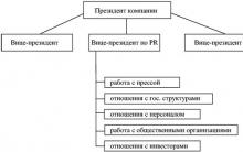
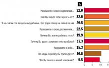

 Future marriage of Prince Harry year NTV")


Energy drinks: give vigor, but take away health What will happen if you drink 4 energy drinks
Mustard for weight loss: how to use the seasoning with maximum benefit Is it possible for children to have mustard
The benefits and harms of mustard for the human body Table mustard benefits and harms
How to treat the ear after piercing: types of antiseptics, their composition, rules and features of the treatment of a pierced ear
Sistine Chapel in the Vatican: description, history, architectural features